L. G. Bobyleva, T. A. Uryupina, N. V. Penkov, M. A. Timchenko, A. D. Ulanova, A. G. Gabdulkhakov, I. M. Vikhlyantsev, A. G. Bobylev
{"title":"The Structural Features of Skeletal Muscle Titin Aggregates","authors":"L. G. Bobyleva, T. A. Uryupina, N. V. Penkov, M. A. Timchenko, A. D. Ulanova, A. G. Gabdulkhakov, I. M. Vikhlyantsev, A. G. Bobylev","doi":"10.1134/s0026893324020043","DOIUrl":null,"url":null,"abstract":"<p><b>Abstract</b>—Titin is a multidomain protein of striated and smooth muscles of vertebrates. The protein consists of repeating immunoglobulin-like (Ig) and fibronectin-like (FnIII) domains, which are β-sandwiches with a predominant β-structure, and also contains disordered regions. In this work, the methods of atomic force microscopy (AFM), X-ray diffraction, and Fourier transform infrared spectroscopy were used to study the morphology and structure of aggregates of rabbit skeletal muscle titin obtained in two different solutions: 0.15 M glycine-KOH, pH 7.0 and 200 mM KCl, 10 mM imidazole, pH 7.0. According to AFM data, skeletal muscle titin formed amorphous aggregates of different morphologies in the above two solutions. Amorphous aggregates of titin formed in a solution containing glycine consisted of much larger particles than aggregates of this protein formed in a solution containing KCl. The “KCl-aggregates” according to AFM data had the form of a “sponge”-like structure, while amorphous “glycine-aggregates” of titin formed “branching” structures. Spectrofluorometry revealed the ability of “glycine-aggregates” of titin to bind to the dye thioflavin T (TT), and X-ray diffraction revealed the presence of one of the elements of the amyloid cross β-structure, a reflection of ~4.6 Å, in these aggregates. These data indicate that “glycine-aggregates” of titin are amyloid or amyloid-like. No similar structural features were found in “KCl-aggregates” of titin; they also did not show the ability to bind to thioflavin T, indicating the non-amyloid nature of these titin aggregates. Fourier transform infrared spectroscopy revealed differences in the secondary structure of the two types of titin aggregates. The data we obtained demonstrate the features of structural changes during the formation of intermolecular bonds between molecules of the giant titin protein during its aggregation. The data expand the understanding of the process of amyloid protein aggregation.</p>","PeriodicalId":18734,"journal":{"name":"Molecular Biology","volume":"44 1","pages":""},"PeriodicalIF":1.2000,"publicationDate":"2024-04-09","publicationTypes":"Journal Article","fieldsOfStudy":null,"isOpenAccess":false,"openAccessPdf":"","citationCount":"0","resultStr":null,"platform":"Semanticscholar","paperid":null,"PeriodicalName":"Molecular Biology","FirstCategoryId":"99","ListUrlMain":"https://doi.org/10.1134/s0026893324020043","RegionNum":4,"RegionCategory":"生物学","ArticlePicture":[],"TitleCN":null,"AbstractTextCN":null,"PMCID":null,"EPubDate":"","PubModel":"","JCR":"Q4","JCRName":"BIOCHEMISTRY & MOLECULAR BIOLOGY","Score":null,"Total":0}
引用次数: 0
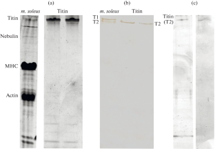
Abstract—Titin is a multidomain protein of striated and smooth muscles of vertebrates. The protein consists of repeating immunoglobulin-like (Ig) and fibronectin-like (FnIII) domains, which are β-sandwiches with a predominant β-structure, and also contains disordered regions. In this work, the methods of atomic force microscopy (AFM), X-ray diffraction, and Fourier transform infrared spectroscopy were used to study the morphology and structure of aggregates of rabbit skeletal muscle titin obtained in two different solutions: 0.15 M glycine-KOH, pH 7.0 and 200 mM KCl, 10 mM imidazole, pH 7.0. According to AFM data, skeletal muscle titin formed amorphous aggregates of different morphologies in the above two solutions. Amorphous aggregates of titin formed in a solution containing glycine consisted of much larger particles than aggregates of this protein formed in a solution containing KCl. The “KCl-aggregates” according to AFM data had the form of a “sponge”-like structure, while amorphous “glycine-aggregates” of titin formed “branching” structures. Spectrofluorometry revealed the ability of “glycine-aggregates” of titin to bind to the dye thioflavin T (TT), and X-ray diffraction revealed the presence of one of the elements of the amyloid cross β-structure, a reflection of ~4.6 Å, in these aggregates. These data indicate that “glycine-aggregates” of titin are amyloid or amyloid-like. No similar structural features were found in “KCl-aggregates” of titin; they also did not show the ability to bind to thioflavin T, indicating the non-amyloid nature of these titin aggregates. Fourier transform infrared spectroscopy revealed differences in the secondary structure of the two types of titin aggregates. The data we obtained demonstrate the features of structural changes during the formation of intermolecular bonds between molecules of the giant titin protein during its aggregation. The data expand the understanding of the process of amyloid protein aggregation.
期刊介绍:
Molecular Biology is an international peer reviewed journal that covers a wide scope of problems in molecular, cell and computational biology including genomics, proteomics, bioinformatics, molecular virology and immunology, molecular development biology, molecular evolution and related areals. Molecular Biology publishes reviews, experimental and theoretical works. Every year, the journal publishes special issues devoted to most rapidly developing branches of physical-chemical biology and to the most outstanding scientists.


 分享
分享
 求助内容:
求助内容: 应助结果提醒方式:
 扫码关注我们
