{"title":"A Novel Structure Fusion Attention Model to Detect Architectural Distortion on Mammography","authors":"Ting-Wei Ou, Tzu-Chieh Weng, Ruey-Feng Chang","doi":"10.1007/s10278-024-01085-y","DOIUrl":null,"url":null,"abstract":"<p>Architectural distortion (AD) is one of the most common findings on mammograms, and it may represent not only cancer but also a lesion such as a radial scar that may have an associated cancer. AD accounts for 18–45% missed cancer, and the positive predictive value of AD is approximately 74.5%. Early detection of AD leads to early diagnosis and treatment of the cancer and improves the overall prognosis. However, detection of AD is a challenging task. In this work, we propose a new approach for detecting architectural distortion in mammography images by combining preprocessing methods and a novel structure fusion attention model. The proposed structure-focused weighted orientation preprocessing method is composed of the original image, the architecture enhancement map, and the weighted orientation map, highlighting suspicious AD locations. The proposed structure fusion attention model captures the information from different channels and outperforms other models in terms of false positives and top sensitivity, which refers to the maximum sensitivity that a model can achieve under the acceptance of the highest number of false positives, reaching 0.92 top sensitivity with only 0.6590 false positive per image. The findings suggest that the combination of preprocessing methods and a novel network architecture can lead to more accurate and reliable AD detection. Overall, the proposed approach offers a novel perspective on detecting ADs, and we believe that our method can be applied to clinical settings in the future, assisting radiologists in the early detection of ADs from mammography, ultimately leading to early treatment of breast cancer patients.</p>","PeriodicalId":50214,"journal":{"name":"Journal of Digital Imaging","volume":"306 1","pages":""},"PeriodicalIF":3.8000,"publicationDate":"2024-04-16","publicationTypes":"Journal Article","fieldsOfStudy":null,"isOpenAccess":false,"openAccessPdf":"","citationCount":"0","resultStr":null,"platform":"Semanticscholar","paperid":null,"PeriodicalName":"Journal of Digital Imaging","FirstCategoryId":"5","ListUrlMain":"https://doi.org/10.1007/s10278-024-01085-y","RegionNum":2,"RegionCategory":"工程技术","ArticlePicture":[],"TitleCN":null,"AbstractTextCN":null,"PMCID":null,"EPubDate":"","PubModel":"","JCR":"Q2","JCRName":"RADIOLOGY, NUCLEAR MEDICINE & MEDICAL IMAGING","Score":null,"Total":0}
引用次数: 0
Abstract
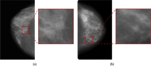
Architectural distortion (AD) is one of the most common findings on mammograms, and it may represent not only cancer but also a lesion such as a radial scar that may have an associated cancer. AD accounts for 18–45% missed cancer, and the positive predictive value of AD is approximately 74.5%. Early detection of AD leads to early diagnosis and treatment of the cancer and improves the overall prognosis. However, detection of AD is a challenging task. In this work, we propose a new approach for detecting architectural distortion in mammography images by combining preprocessing methods and a novel structure fusion attention model. The proposed structure-focused weighted orientation preprocessing method is composed of the original image, the architecture enhancement map, and the weighted orientation map, highlighting suspicious AD locations. The proposed structure fusion attention model captures the information from different channels and outperforms other models in terms of false positives and top sensitivity, which refers to the maximum sensitivity that a model can achieve under the acceptance of the highest number of false positives, reaching 0.92 top sensitivity with only 0.6590 false positive per image. The findings suggest that the combination of preprocessing methods and a novel network architecture can lead to more accurate and reliable AD detection. Overall, the proposed approach offers a novel perspective on detecting ADs, and we believe that our method can be applied to clinical settings in the future, assisting radiologists in the early detection of ADs from mammography, ultimately leading to early treatment of breast cancer patients.
建筑变形(AD)是乳房 X 光检查中最常见的发现之一,它不仅可能代表癌症,也可能代表可能伴有癌症的病变,如放射状疤痕。AD占漏诊癌症的18%-45%,AD的阳性预测值约为74.5%。早期发现 AD 可使癌症得到早期诊断和治疗,并改善整体预后。然而,检测 AD 是一项具有挑战性的任务。在这项工作中,我们结合预处理方法和新型结构融合注意力模型,提出了一种检测乳腺 X 射线图像结构失真的新方法。所提出的以结构为重点的加权方向预处理方法由原始图像、结构增强图和加权方向图组成,可突出显示可疑的乳腺增生位置。所提出的结构融合注意力模型捕捉了来自不同通道的信息,在误报率和最高灵敏度(指模型在接受最高误报率的情况下所能达到的最高灵敏度)方面优于其他模型,最高灵敏度达到 0.92,而每幅图像的误报率仅为 0.6590。研究结果表明,将预处理方法与新型网络架构相结合,可以实现更准确、更可靠的注意力缺失检测。总之,所提出的方法为检测乳腺增生症提供了一个新的视角,我们相信我们的方法将来可以应用于临床,帮助放射科医生从乳腺 X 射线摄影中早期检测出乳腺增生症,最终实现乳腺癌患者的早期治疗。
期刊介绍:
The Journal of Digital Imaging (JDI) is the official peer-reviewed journal of the Society for Imaging Informatics in Medicine (SIIM). JDI’s goal is to enhance the exchange of knowledge encompassed by the general topic of Imaging Informatics in Medicine such as research and practice in clinical, engineering, and information technologies and techniques in all medical imaging environments. JDI topics are of interest to researchers, developers, educators, physicians, and imaging informatics professionals.
Suggested Topics
PACS and component systems; imaging informatics for the enterprise; image-enabled electronic medical records; RIS and HIS; digital image acquisition; image processing; image data compression; 3D, visualization, and multimedia; speech recognition; computer-aided diagnosis; facilities design; imaging vocabularies and ontologies; Transforming the Radiological Interpretation Process (TRIP™); DICOM and other standards; workflow and process modeling and simulation; quality assurance; archive integrity and security; teleradiology; digital mammography; and radiological informatics education.


 分享
分享
 求助内容:
求助内容: 应助结果提醒方式:
 扫码关注我们
