{"title":"Personalized voltage maps guided by cardiac magnetic resonance in the era of high-density mapping","authors":"","doi":"10.1016/j.hrthm.2024.04.074","DOIUrl":null,"url":null,"abstract":"<div><h3>Background</h3><div>Voltage mapping could identify the conducting channels potentially responsible for ventricular tachycardia (VT). Standard thresholds (0.5–1.5 mV) were established using bipolar catheters. No thresholds have been analyzed with high-density mapping catheters. In addition, channels identified by cardiac magnetic resonance (CMR) has been proven to be related with VT.</div></div><div><h3>Objective</h3><div>The purpose of this study was to analyze the diagnostic yield of a personalized voltage map using CMR to guide the adjustment of voltage thresholds.</div></div><div><h3>Methods</h3><div>All consecutive patients with scar-related VT undergoing ablation after CMR (from October 2018 to December 2020) were included. First, personalized CMR-guided voltage thresholds were defined systematically according to the distribution of the scar and channels. Second, to validate these new thresholds, a comparison with standard thresholds (0.5–1.5 mV) was performed. Tissue characteristics of areas identified as deceleration zones (DZs) were recorded for each pair of thresholds. In addition, the relation of VT circuits with voltage channels was analyzed for both maps.</div></div><div><h3>Results</h3><div>Thirty-two patients were included [mean age 66.6 ± 11.2 years; 25 (78.1%) ischemic cardiomyopathy]. Overall, 52 DZs were observed: 44.2% were identified as border zone tissue with standard cutoffs vs 75.0% using personalized voltage thresholds (<em>P</em> = .003). Of the 31 VT isthmuses detected, only 35.5% correlated with a voltage channel with standard thresholds vs 74.2% using adjusted thresholds (<em>P</em> = .005). Adjusted cutoff bipolar voltages that better matched CMR images were 0.51 ± 0.32 and 1.79 ± 0.71 mV with high interindividual variability (from 0.14–1.68 to 0.7–3.21 mV).</div></div><div><h3>Conclusion</h3><div>Personalized voltage CMR-guided personalized voltage maps enable a better identification of the substrate with a higher correlation with both DZs and VT isthmuses than do conventional voltage maps using fixed thresholds.</div></div>","PeriodicalId":12886,"journal":{"name":"Heart rhythm","volume":"21 10","pages":"Pages 1811-1819"},"PeriodicalIF":5.7000,"publicationDate":"2024-10-01","publicationTypes":"Journal Article","fieldsOfStudy":null,"isOpenAccess":false,"openAccessPdf":"","citationCount":"0","resultStr":null,"platform":"Semanticscholar","paperid":null,"PeriodicalName":"Heart rhythm","FirstCategoryId":"3","ListUrlMain":"https://www.sciencedirect.com/science/article/pii/S1547527124025013","RegionNum":2,"RegionCategory":"医学","ArticlePicture":[],"TitleCN":null,"AbstractTextCN":null,"PMCID":null,"EPubDate":"","PubModel":"","JCR":"Q1","JCRName":"CARDIAC & CARDIOVASCULAR SYSTEMS","Score":null,"Total":0}
引用次数: 0
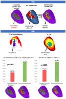
Abstract
Background
Voltage mapping could identify the conducting channels potentially responsible for ventricular tachycardia (VT). Standard thresholds (0.5–1.5 mV) were established using bipolar catheters. No thresholds have been analyzed with high-density mapping catheters. In addition, channels identified by cardiac magnetic resonance (CMR) has been proven to be related with VT.
Objective
The purpose of this study was to analyze the diagnostic yield of a personalized voltage map using CMR to guide the adjustment of voltage thresholds.
Methods
All consecutive patients with scar-related VT undergoing ablation after CMR (from October 2018 to December 2020) were included. First, personalized CMR-guided voltage thresholds were defined systematically according to the distribution of the scar and channels. Second, to validate these new thresholds, a comparison with standard thresholds (0.5–1.5 mV) was performed. Tissue characteristics of areas identified as deceleration zones (DZs) were recorded for each pair of thresholds. In addition, the relation of VT circuits with voltage channels was analyzed for both maps.
Results
Thirty-two patients were included [mean age 66.6 ± 11.2 years; 25 (78.1%) ischemic cardiomyopathy]. Overall, 52 DZs were observed: 44.2% were identified as border zone tissue with standard cutoffs vs 75.0% using personalized voltage thresholds (P = .003). Of the 31 VT isthmuses detected, only 35.5% correlated with a voltage channel with standard thresholds vs 74.2% using adjusted thresholds (P = .005). Adjusted cutoff bipolar voltages that better matched CMR images were 0.51 ± 0.32 and 1.79 ± 0.71 mV with high interindividual variability (from 0.14–1.68 to 0.7–3.21 mV).
Conclusion
Personalized voltage CMR-guided personalized voltage maps enable a better identification of the substrate with a higher correlation with both DZs and VT isthmuses than do conventional voltage maps using fixed thresholds.
期刊介绍:
HeartRhythm, the official Journal of the Heart Rhythm Society and the Cardiac Electrophysiology Society, is a unique journal for fundamental discovery and clinical applicability.
HeartRhythm integrates the entire cardiac electrophysiology (EP) community from basic and clinical academic researchers, private practitioners, engineers, allied professionals, industry, and trainees, all of whom are vital and interdependent members of our EP community.
The Heart Rhythm Society is the international leader in science, education, and advocacy for cardiac arrhythmia professionals and patients, and the primary information resource on heart rhythm disorders. Its mission is to improve the care of patients by promoting research, education, and optimal health care policies and standards.


 分享
分享
 求助内容:
求助内容: 应助结果提醒方式:
 扫码关注我们
