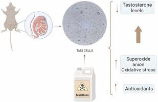
{"title":"Lower malathion concentrations reduce testosterone biosynthesis by Leydig TM3 cells in vitro by altering cellular redox profile and inducing oxidative damage","authors":"Rafaela Pires Erthal-Michelato , Débora Hipólito Quadreli , Tiago Henrique Zaninelli , Waldiceu Aparecido Verri , Glaura Scantamburlo Alves Fernandes","doi":"10.1016/j.reprotox.2024.108595","DOIUrl":null,"url":null,"abstract":"<div><p>Malathion is an organophosphate pesticide used in agriculture and control of the <em>Aedes aegypti</em> mosquito. As previous reports have indicated the potential of malathion to compromise testosterone production in in vivo models, the objective of this study was to elucidate the mechanisms underlying the impairment of Leydig cell function, considering its critical role in male reproductive function. To this end, murine Leydig TM3 cells were exposed to concentrations of 1, 10, 100 or 1000 μM malathion for 24 h for evaluation of the compound on cell viability. Subsequently, concentrations of 1, 10, and 100 μM malathion were employed for a 24-h period to assess testosterone biosynthesis, levels of cytokines IL-1β, IL-6, IL-10, and TNF-α, as well as the redox profile. Malathion exerted a concentration-dependent impact on cell viability. Notably, the lower concentrations of malathion (1 and 10 μM) were found to impair testosterone biosynthesis in TM3 cells. While there were changes in IL-1 and TNF-α levels at specific concentrations, no direct correlation with altered hormone production was established. Our investigation revealed that varied malathion concentrations induced oxidative stress by increase in superoxide anion and a compensatory rise in antioxidants. In conclusion, the observed changes in the oxidative profile of TM3 cells were linked to functional impairment, evidenced by reduced testosterone biosynthesis at lower malathion concentrations.</p></div>","PeriodicalId":21137,"journal":{"name":"Reproductive toxicology","volume":"126 ","pages":"Article 108595"},"PeriodicalIF":2.8000,"publicationDate":"2024-04-17","publicationTypes":"Journal Article","fieldsOfStudy":null,"isOpenAccess":false,"openAccessPdf":"","citationCount":"0","resultStr":null,"platform":"Semanticscholar","paperid":null,"PeriodicalName":"Reproductive toxicology","FirstCategoryId":"3","ListUrlMain":"https://www.sciencedirect.com/science/article/pii/S0890623824000625","RegionNum":4,"RegionCategory":"医学","ArticlePicture":[],"TitleCN":null,"AbstractTextCN":null,"PMCID":null,"EPubDate":"","PubModel":"","JCR":"Q2","JCRName":"REPRODUCTIVE BIOLOGY","Score":null,"Total":0}
引用次数: 0
Abstract
Malathion is an organophosphate pesticide used in agriculture and control of the Aedes aegypti mosquito. As previous reports have indicated the potential of malathion to compromise testosterone production in in vivo models, the objective of this study was to elucidate the mechanisms underlying the impairment of Leydig cell function, considering its critical role in male reproductive function. To this end, murine Leydig TM3 cells were exposed to concentrations of 1, 10, 100 or 1000 μM malathion for 24 h for evaluation of the compound on cell viability. Subsequently, concentrations of 1, 10, and 100 μM malathion were employed for a 24-h period to assess testosterone biosynthesis, levels of cytokines IL-1β, IL-6, IL-10, and TNF-α, as well as the redox profile. Malathion exerted a concentration-dependent impact on cell viability. Notably, the lower concentrations of malathion (1 and 10 μM) were found to impair testosterone biosynthesis in TM3 cells. While there were changes in IL-1 and TNF-α levels at specific concentrations, no direct correlation with altered hormone production was established. Our investigation revealed that varied malathion concentrations induced oxidative stress by increase in superoxide anion and a compensatory rise in antioxidants. In conclusion, the observed changes in the oxidative profile of TM3 cells were linked to functional impairment, evidenced by reduced testosterone biosynthesis at lower malathion concentrations.
期刊介绍:
Drawing from a large number of disciplines, Reproductive Toxicology publishes timely, original research on the influence of chemical and physical agents on reproduction. Written by and for obstetricians, pediatricians, embryologists, teratologists, geneticists, toxicologists, andrologists, and others interested in detecting potential reproductive hazards, the journal is a forum for communication among researchers and practitioners. Articles focus on the application of in vitro, animal and clinical research to the practice of clinical medicine.
All aspects of reproduction are within the scope of Reproductive Toxicology, including the formation and maturation of male and female gametes, sexual function, the events surrounding the fusion of gametes and the development of the fertilized ovum, nourishment and transport of the conceptus within the genital tract, implantation, embryogenesis, intrauterine growth, placentation and placental function, parturition, lactation and neonatal survival. Adverse reproductive effects in males will be considered as significant as adverse effects occurring in females. To provide a balanced presentation of approaches, equal emphasis will be given to clinical and animal or in vitro work. Typical end points that will be studied by contributors include infertility, sexual dysfunction, spontaneous abortion, malformations, abnormal histogenesis, stillbirth, intrauterine growth retardation, prematurity, behavioral abnormalities, and perinatal mortality.


 分享
分享
 求助内容:
求助内容: 应助结果提醒方式:
 扫码关注我们
