Z. Guo , D.P. Guillen , J.R. Grimm , C. Renteria , C. Marsico , V. Nikitin , D. Arola
{"title":"High throughput automated characterization of enamel microstructure using synchrotron tomography and optical flow imaging","authors":"Z. Guo , D.P. Guillen , J.R. Grimm , C. Renteria , C. Marsico , V. Nikitin , D. Arola","doi":"10.1016/j.actbio.2024.04.033","DOIUrl":null,"url":null,"abstract":"<div><p>The remarkable damage-tolerance of enamel has been attributed to its hierarchical microstructure and the organized bands of decussated rods. A thorough characterization of the microscale rod evolution within the enamel is needed to elucidate this complex structure. While prior efforts in this area have made use of single particle tracking to track a single rod evolution to various degrees of success, such a process can be both computationally and labor intensive, limited to the evolution path of a single rod, and is therefore prone to error from potentially tracking outliers. Particle image velocimetry (PIV) is a well-established algorithm to derive field information from image sequences for processes that are time-dependent, such as fluid flows and structural deformation. In this work, we demonstrate the use of PIV in extracting the full-field microstructural distribution of rods within the enamel. Enamel samples from a wild African lion were analyzed using high-energy synchrotron X-ray micro-tomography. Results from the PIV analysis provide sufficient full-field information to reconstruct the growth of individual rods that can potentially enable rapid analysis of complex microstructures from high resolution synchrotron datasets. Such information can serve as a template for designing damage-tolerant bioinspired structures for advanced manufacturing.</p></div><div><h3>Statement of significance</h3><p>Thorough characterization and analysis of biological microstructures (viz. dental enamel) allows us to understand the basis of their excellent mechanical properties. Prior efforts have successfully replicated these microstructures via single particle tracking, but the process is computationally and labor intensive. In this work, optical flow imaging algorithms were used to extract full-field microstructural distribution of enamel rods from synchrotron X-ray computed tomography datasets, and a field method was used to reconstruct the growth of individual rods. Such high throughput information allows for the rapid production/prototyping and advanced manufacturing of damage-tolerant bioinspired structures for specific engineering applications. Furthermore, the algorithms used herein are freely available and open source to broaden the availability of the proposed workflow to the general scientific community.</p></div>","PeriodicalId":237,"journal":{"name":"Acta Biomaterialia","volume":"181 ","pages":"Pages 263-271"},"PeriodicalIF":9.6000,"publicationDate":"2024-06-01","publicationTypes":"Journal Article","fieldsOfStudy":null,"isOpenAccess":false,"openAccessPdf":"","citationCount":"0","resultStr":null,"platform":"Semanticscholar","paperid":null,"PeriodicalName":"Acta Biomaterialia","FirstCategoryId":"5","ListUrlMain":"https://www.sciencedirect.com/science/article/pii/S1742706124002162","RegionNum":1,"RegionCategory":"医学","ArticlePicture":[],"TitleCN":null,"AbstractTextCN":null,"PMCID":null,"EPubDate":"2024/4/25 0:00:00","PubModel":"Epub","JCR":"Q1","JCRName":"ENGINEERING, BIOMEDICAL","Score":null,"Total":0}
引用次数: 0
Abstract
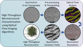
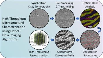
The remarkable damage-tolerance of enamel has been attributed to its hierarchical microstructure and the organized bands of decussated rods. A thorough characterization of the microscale rod evolution within the enamel is needed to elucidate this complex structure. While prior efforts in this area have made use of single particle tracking to track a single rod evolution to various degrees of success, such a process can be both computationally and labor intensive, limited to the evolution path of a single rod, and is therefore prone to error from potentially tracking outliers. Particle image velocimetry (PIV) is a well-established algorithm to derive field information from image sequences for processes that are time-dependent, such as fluid flows and structural deformation. In this work, we demonstrate the use of PIV in extracting the full-field microstructural distribution of rods within the enamel. Enamel samples from a wild African lion were analyzed using high-energy synchrotron X-ray micro-tomography. Results from the PIV analysis provide sufficient full-field information to reconstruct the growth of individual rods that can potentially enable rapid analysis of complex microstructures from high resolution synchrotron datasets. Such information can serve as a template for designing damage-tolerant bioinspired structures for advanced manufacturing.
Statement of significance
Thorough characterization and analysis of biological microstructures (viz. dental enamel) allows us to understand the basis of their excellent mechanical properties. Prior efforts have successfully replicated these microstructures via single particle tracking, but the process is computationally and labor intensive. In this work, optical flow imaging algorithms were used to extract full-field microstructural distribution of enamel rods from synchrotron X-ray computed tomography datasets, and a field method was used to reconstruct the growth of individual rods. Such high throughput information allows for the rapid production/prototyping and advanced manufacturing of damage-tolerant bioinspired structures for specific engineering applications. Furthermore, the algorithms used herein are freely available and open source to broaden the availability of the proposed workflow to the general scientific community.
珐琅质卓越的耐损伤性归功于其层次分明的微观结构和有组织的簇状棒带。为了阐明这种复杂的结构,需要对珐琅质内部微观尺度的杆演变进行彻底的表征。虽然之前在这一领域的研究在不同程度上成功地利用了单颗粒跟踪技术来跟踪单个杆的演变,但这种方法既耗费计算量又耗费人力,而且仅限于单个杆的演变路径,因此很容易因可能跟踪到异常值而产生误差。粒子图像测速(PIV)是一种成熟的算法,可从图像序列中获取与时间相关的过程(如流体流动和结构变形)的现场信息。在这项工作中,我们展示了如何利用粒子测距法提取珐琅质内棒状物的全场微观结构分布。我们使用高能同步辐射 X 射线显微层析技术分析了非洲野生狮子的珐琅质样本。PIV 分析结果提供了足够的全场信息,可以重建单个棒状物的生长过程,从而有可能从高分辨率同步辐射数据集中快速分析复杂的微观结构。对生物微结构(如牙釉质)进行彻底的表征和分析,可让我们了解其优异机械性能的基础。之前的研究已通过单颗粒跟踪成功复制了这些微结构,但这一过程需要大量的计算和人力。在这项工作中,我们使用光流成像算法从同步辐射 X 射线计算机断层扫描数据集中提取珐琅质棒的全场微结构分布,并使用场方法重建单个棒的生长过程。通过这种高通量信息,可以为特定工程应用快速生产/原型开发和先进制造耐损伤生物启发结构。此外,本文所使用的算法是免费提供和开源的,以扩大拟议工作流程对广大科学界的可用性。
期刊介绍:
Acta Biomaterialia is a monthly peer-reviewed scientific journal published by Elsevier. The journal was established in January 2005. The editor-in-chief is W.R. Wagner (University of Pittsburgh). The journal covers research in biomaterials science, including the interrelationship of biomaterial structure and function from macroscale to nanoscale. Topical coverage includes biomedical and biocompatible materials.


 分享
分享
 求助内容:
求助内容: 应助结果提醒方式:
 扫码关注我们
