Mitchell Josvai , Erzsebet Polyak , Meghana Kalluri , Samantha Robertson , Wendy C. Crone , Masatoshi Suzuki
{"title":"An engineered in vitro model of the human myotendinous junction","authors":"Mitchell Josvai , Erzsebet Polyak , Meghana Kalluri , Samantha Robertson , Wendy C. Crone , Masatoshi Suzuki","doi":"10.1016/j.actbio.2024.04.007","DOIUrl":null,"url":null,"abstract":"<div><p>The myotendinous junction (MTJ) is a vulnerable region at the interface of skeletal muscle and tendon that forms an integrated mechanical unit. This study presents a technique for the spatially restrictive co-culture of human embryonic stem cell (hESC)-derived skeletal myocytes and primary tenocytes for two-dimensional modeling of the MTJ. Micropatterned lanes of extracellular matrix and a 2-well culture chamber define the initial regions of occupation. On day 1, both lines occupy less than 20 % of the initially vacant interstitial zone, referred to henceforth as the junction. Myocyte–tenocyte interdigitations are observed by day 7. Immunocytochemistry reveals enhanced organization and alignment of patterned myocyte and tenocyte features, as well as differential expression of multiple MTJ markers. On day 24, electrically stimulated junction myocytes demonstrate negative contractile strains, while positive tensile strains are exhibited by mechanically passive tenocytes at the junction. Unpatterned tenocytes distal to the junction experience significantly decreased strains in comparison to cells at the interface. Unpatterned myocytes have impaired organization and uncoordinated contractile behavior. These findings suggest that this platform is capable of inducing myocyte–tenocyte junction formation and mechanical coupling similar to the native MTJ, showing transduction of force across the cell–cell interface.</p></div><div><h3>Statement of significance</h3><p>The myotendinous junction (MTJ) is an integrated structure that transduces force across the muscle-tendon boundary, making the region vulnerable to strain injury. Despite the clinical relevance, previous <em>in vitro</em> models of the MTJ lack the structure and mechanical accuracy of the native tissue and have difficulty transmitting force across the cell–cell interface. This study demonstrates an <em>in vitro</em> model of the MTJ, using spatially restrictive cues to inform human myocyte–tenocyte interactions and architecture. The model expressed MTJ markers and developed anisotropic myocyte–tenocyte integrations that resemble the native tissue and allow for force transduction from contracting myocytes to passive tenocyte regions. As such, this study presents a system capable of investigating development, injury, and pathology in the human MTJ.</p></div>","PeriodicalId":237,"journal":{"name":"Acta Biomaterialia","volume":"180 ","pages":"Pages 279-294"},"PeriodicalIF":9.6000,"publicationDate":"2024-05-01","publicationTypes":"Journal Article","fieldsOfStudy":null,"isOpenAccess":false,"openAccessPdf":"","citationCount":"0","resultStr":null,"platform":"Semanticscholar","paperid":null,"PeriodicalName":"Acta Biomaterialia","FirstCategoryId":"5","ListUrlMain":"https://www.sciencedirect.com/science/article/pii/S1742706124001806","RegionNum":1,"RegionCategory":"医学","ArticlePicture":[],"TitleCN":null,"AbstractTextCN":null,"PMCID":null,"EPubDate":"","PubModel":"","JCR":"Q1","JCRName":"ENGINEERING, BIOMEDICAL","Score":null,"Total":0}
引用次数: 0
Abstract
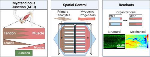
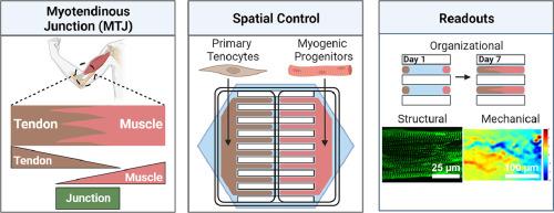
The myotendinous junction (MTJ) is a vulnerable region at the interface of skeletal muscle and tendon that forms an integrated mechanical unit. This study presents a technique for the spatially restrictive co-culture of human embryonic stem cell (hESC)-derived skeletal myocytes and primary tenocytes for two-dimensional modeling of the MTJ. Micropatterned lanes of extracellular matrix and a 2-well culture chamber define the initial regions of occupation. On day 1, both lines occupy less than 20 % of the initially vacant interstitial zone, referred to henceforth as the junction. Myocyte–tenocyte interdigitations are observed by day 7. Immunocytochemistry reveals enhanced organization and alignment of patterned myocyte and tenocyte features, as well as differential expression of multiple MTJ markers. On day 24, electrically stimulated junction myocytes demonstrate negative contractile strains, while positive tensile strains are exhibited by mechanically passive tenocytes at the junction. Unpatterned tenocytes distal to the junction experience significantly decreased strains in comparison to cells at the interface. Unpatterned myocytes have impaired organization and uncoordinated contractile behavior. These findings suggest that this platform is capable of inducing myocyte–tenocyte junction formation and mechanical coupling similar to the native MTJ, showing transduction of force across the cell–cell interface.
Statement of significance
The myotendinous junction (MTJ) is an integrated structure that transduces force across the muscle-tendon boundary, making the region vulnerable to strain injury. Despite the clinical relevance, previous in vitro models of the MTJ lack the structure and mechanical accuracy of the native tissue and have difficulty transmitting force across the cell–cell interface. This study demonstrates an in vitro model of the MTJ, using spatially restrictive cues to inform human myocyte–tenocyte interactions and architecture. The model expressed MTJ markers and developed anisotropic myocyte–tenocyte integrations that resemble the native tissue and allow for force transduction from contracting myocytes to passive tenocyte regions. As such, this study presents a system capable of investigating development, injury, and pathology in the human MTJ.
期刊介绍:
Acta Biomaterialia is a monthly peer-reviewed scientific journal published by Elsevier. The journal was established in January 2005. The editor-in-chief is W.R. Wagner (University of Pittsburgh). The journal covers research in biomaterials science, including the interrelationship of biomaterial structure and function from macroscale to nanoscale. Topical coverage includes biomedical and biocompatible materials.


 分享
分享
 求助内容:
求助内容: 应助结果提醒方式:
 扫码关注我们
