Super-resolution Deep Learning Reconstruction Cervical Spine 1.5T MRI: Improved Interobserver Agreement in Evaluations of Neuroforaminal Stenosis Compared to Conventional Deep Learning Reconstruction
{"title":"Super-resolution Deep Learning Reconstruction Cervical Spine 1.5T MRI: Improved Interobserver Agreement in Evaluations of Neuroforaminal Stenosis Compared to Conventional Deep Learning Reconstruction","authors":"Koichiro Yasaka, Shunichi Uehara, Shimpei Kato, Yusuke Watanabe, Taku Tajima, Hiroyuki Akai, Naoki Yoshioka, Masaaki Akahane, Kuni Ohtomo, Osamu Abe, Shigeru Kiryu","doi":"10.1007/s10278-024-01112-y","DOIUrl":null,"url":null,"abstract":"<p>The aim of this study was to investigate whether super-resolution deep learning reconstruction (SR-DLR) is superior to conventional deep learning reconstruction (DLR) with respect to interobserver agreement in the evaluation of neuroforaminal stenosis using 1.5T cervical spine MRI. This retrospective study included 39 patients who underwent 1.5T cervical spine MRI. T2-weighted sagittal images were reconstructed with SR-DLR and DLR. Three blinded radiologists independently evaluated the images in terms of the degree of neuroforaminal stenosis, depictions of the vertebrae, spinal cord and neural foramina, sharpness, noise, artefacts and diagnostic acceptability. In quantitative image analyses, a fourth radiologist evaluated the signal-to-noise ratio (SNR) by placing a circular or ovoid region of interest on the spinal cord, and the edge slope based on a linear region of interest placed across the surface of the spinal cord. Interobserver agreement in the evaluations of neuroforaminal stenosis using SR-DLR and DLR was 0.422–0.571 and 0.410–0.542, respectively. The kappa values between reader 1 vs. reader 2 and reader 2 vs. reader 3 significantly differed. Two of the three readers rated depictions of the spinal cord, sharpness, and diagnostic acceptability as significantly better with SR-DLR than with DLR. Both SNR and edge slope (/mm) were also significantly better with SR-DLR (12.9 and 6031, respectively) than with DLR (11.5 and 3741, respectively) (<i>p</i> < 0.001 for both). In conclusion, compared to DLR, SR-DLR improved interobserver agreement in the evaluations of neuroforaminal stenosis using 1.5T cervical spine MRI.</p>","PeriodicalId":50214,"journal":{"name":"Journal of Digital Imaging","volume":"09 1","pages":""},"PeriodicalIF":3.8000,"publicationDate":"2024-04-26","publicationTypes":"Journal Article","fieldsOfStudy":null,"isOpenAccess":false,"openAccessPdf":"","citationCount":"0","resultStr":null,"platform":"Semanticscholar","paperid":null,"PeriodicalName":"Journal of Digital Imaging","FirstCategoryId":"5","ListUrlMain":"https://doi.org/10.1007/s10278-024-01112-y","RegionNum":2,"RegionCategory":"工程技术","ArticlePicture":[],"TitleCN":null,"AbstractTextCN":null,"PMCID":null,"EPubDate":"","PubModel":"","JCR":"Q2","JCRName":"RADIOLOGY, NUCLEAR MEDICINE & MEDICAL IMAGING","Score":null,"Total":0}
引用次数: 0
Abstract
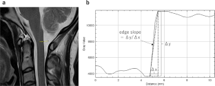
The aim of this study was to investigate whether super-resolution deep learning reconstruction (SR-DLR) is superior to conventional deep learning reconstruction (DLR) with respect to interobserver agreement in the evaluation of neuroforaminal stenosis using 1.5T cervical spine MRI. This retrospective study included 39 patients who underwent 1.5T cervical spine MRI. T2-weighted sagittal images were reconstructed with SR-DLR and DLR. Three blinded radiologists independently evaluated the images in terms of the degree of neuroforaminal stenosis, depictions of the vertebrae, spinal cord and neural foramina, sharpness, noise, artefacts and diagnostic acceptability. In quantitative image analyses, a fourth radiologist evaluated the signal-to-noise ratio (SNR) by placing a circular or ovoid region of interest on the spinal cord, and the edge slope based on a linear region of interest placed across the surface of the spinal cord. Interobserver agreement in the evaluations of neuroforaminal stenosis using SR-DLR and DLR was 0.422–0.571 and 0.410–0.542, respectively. The kappa values between reader 1 vs. reader 2 and reader 2 vs. reader 3 significantly differed. Two of the three readers rated depictions of the spinal cord, sharpness, and diagnostic acceptability as significantly better with SR-DLR than with DLR. Both SNR and edge slope (/mm) were also significantly better with SR-DLR (12.9 and 6031, respectively) than with DLR (11.5 and 3741, respectively) (p < 0.001 for both). In conclusion, compared to DLR, SR-DLR improved interobserver agreement in the evaluations of neuroforaminal stenosis using 1.5T cervical spine MRI.
期刊介绍:
The Journal of Digital Imaging (JDI) is the official peer-reviewed journal of the Society for Imaging Informatics in Medicine (SIIM). JDI’s goal is to enhance the exchange of knowledge encompassed by the general topic of Imaging Informatics in Medicine such as research and practice in clinical, engineering, and information technologies and techniques in all medical imaging environments. JDI topics are of interest to researchers, developers, educators, physicians, and imaging informatics professionals.
Suggested Topics
PACS and component systems; imaging informatics for the enterprise; image-enabled electronic medical records; RIS and HIS; digital image acquisition; image processing; image data compression; 3D, visualization, and multimedia; speech recognition; computer-aided diagnosis; facilities design; imaging vocabularies and ontologies; Transforming the Radiological Interpretation Process (TRIP™); DICOM and other standards; workflow and process modeling and simulation; quality assurance; archive integrity and security; teleradiology; digital mammography; and radiological informatics education.


 分享
分享
 求助内容:
求助内容: 应助结果提醒方式:
 扫码关注我们
