Flow cytometric immunophenotypic differentiation patterns of bone marrow eosinophilopoiesis

IF 2.3 3区 医学 Q3 MEDICAL LABORATORY TECHNOLOGY Cytometry Part B: Clinical Cytometry Pub Date : 2024-04-26 DOI:10.1002/cyto.b.22174
Christopher J. Trindade, Xiaoping Sun, Dragan Maric, Sachein Sharma, Hirsh D. Komarow, Christopher S. Hourigan, Amy Klion, Irina Maric
{"title":"Flow cytometric immunophenotypic differentiation patterns of bone marrow eosinophilopoiesis","authors":"Christopher J. Trindade,&nbsp;Xiaoping Sun,&nbsp;Dragan Maric,&nbsp;Sachein Sharma,&nbsp;Hirsh D. Komarow,&nbsp;Christopher S. Hourigan,&nbsp;Amy Klion,&nbsp;Irina Maric","doi":"10.1002/cyto.b.22174","DOIUrl":null,"url":null,"abstract":"<div>\n \n \n <section>\n \n <h3> Background</h3>\n \n <p>Flow cytometry has been widely used to study immunophenotypic patterns of maturation of most hematopoietic lineages in normal human bone marrow aspirates, thus allowing identification of changes in patterns in many myeloid malignancies. Eosinophils play an important role in a wide variety of disorders, including some myeloid neoplasms. However, changes in flow cytometric immunophenotypic patterns during normal and abnormal bone marrow eosinophilopoiesis have not been well studied.</p>\n </section>\n \n <section>\n \n <h3> Methods</h3>\n \n <p>Fresh bone marrow aspirates from 15 healthy donors, 19 patients with hypereosinophilic syndromes (HES), and 11 patients with systemic mastocytosis (SM) were analyzed for candidate markers that included EMR-1, Siglec-8, CCR3, CD9, CD11a, CD11b, CD11c, CD13, CD16, CD29, CD34, CD38, CD45, CD44, CD49d, CD49f, CD54, CD62L, CD69, CD117, CD125 (IL-5Rα), HLA-DR, using 10 parameter flow cytometry. Putative CD34-negative immature and mature normal eosinophil populations were first identified based on changes in expression of the above markers in healthy donors, then confirmed using fluorescence-based cell sorting and morphological evaluation of cytospin preparations. The normal immunophenotypic patterns were then compared to immunophenotypic patterns of eosinophilopoiesis in patients with HES and SM.</p>\n </section>\n \n <section>\n \n <h3> Results</h3>\n \n <p>The eosinophilic lineage was first verified using the human eosinophil-specific antibody EMR-1 in combination with anti-IL-5Rα antibody. Then, a combination of Siglec-8, CD9, CD11b, CCR3, CD49d, and CD49f antibodies was used to delineate normal eosinophilic maturational patterns. Early stages (eosinophilic promyelocytes/myelocytes) were identified as Siglec-8 dim/CD11b dim to moderate/CD9 dim/CCR3 dim/CD49d bright/CD49f dim, intermediate stages (eosinophilic myelocytes/metamyelocytes) as Siglec-8 moderate/CD11b moderate to bright/CD9 moderate/CCR3 moderate/CD49d moderate/CD49f moderate and mature bands/segmented eosinophils as Siglec-8 bright/CD11b bright/CD9 bright/CCR3 bright/CD49d dim/CD49f bright. Overall maturational patterns were also similar in patients with HES and SM; however, the expression levels of several surface markers were altered compared to normal eosinophils.</p>\n </section>\n \n <section>\n \n <h3> Conclusion</h3>\n \n <p>A novel flow cytometric antibody panel was devised to detect alterations in immunophenotypic patterns of bone marrow eosinophil maturation and evaluated in normal, HES and SM samples. This approach will allow us to elucidate changes in immunophenotypic patterns of bone marrow eosinophilopoiesis in other hematological diseases.</p>\n </section>\n </div>","PeriodicalId":10883,"journal":{"name":"Cytometry Part B: Clinical Cytometry","volume":"106 5","pages":"370-382"},"PeriodicalIF":2.3000,"publicationDate":"2024-04-26","publicationTypes":"Journal Article","fieldsOfStudy":null,"isOpenAccess":false,"openAccessPdf":"https://onlinelibrary.wiley.com/doi/epdf/10.1002/cyto.b.22174","citationCount":"0","resultStr":null,"platform":"Semanticscholar","paperid":null,"PeriodicalName":"Cytometry Part B: Clinical Cytometry","FirstCategoryId":"3","ListUrlMain":"https://onlinelibrary.wiley.com/doi/10.1002/cyto.b.22174","RegionNum":3,"RegionCategory":"医学","ArticlePicture":[],"TitleCN":null,"AbstractTextCN":null,"PMCID":null,"EPubDate":"","PubModel":"","JCR":"Q3","JCRName":"MEDICAL LABORATORY TECHNOLOGY","Score":null,"Total":0}
引用次数: 0

Abstract

Background

Flow cytometry has been widely used to study immunophenotypic patterns of maturation of most hematopoietic lineages in normal human bone marrow aspirates, thus allowing identification of changes in patterns in many myeloid malignancies. Eosinophils play an important role in a wide variety of disorders, including some myeloid neoplasms. However, changes in flow cytometric immunophenotypic patterns during normal and abnormal bone marrow eosinophilopoiesis have not been well studied.

Methods

Fresh bone marrow aspirates from 15 healthy donors, 19 patients with hypereosinophilic syndromes (HES), and 11 patients with systemic mastocytosis (SM) were analyzed for candidate markers that included EMR-1, Siglec-8, CCR3, CD9, CD11a, CD11b, CD11c, CD13, CD16, CD29, CD34, CD38, CD45, CD44, CD49d, CD49f, CD54, CD62L, CD69, CD117, CD125 (IL-5Rα), HLA-DR, using 10 parameter flow cytometry. Putative CD34-negative immature and mature normal eosinophil populations were first identified based on changes in expression of the above markers in healthy donors, then confirmed using fluorescence-based cell sorting and morphological evaluation of cytospin preparations. The normal immunophenotypic patterns were then compared to immunophenotypic patterns of eosinophilopoiesis in patients with HES and SM.

Results

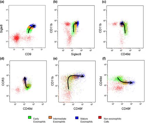
The eosinophilic lineage was first verified using the human eosinophil-specific antibody EMR-1 in combination with anti-IL-5Rα antibody. Then, a combination of Siglec-8, CD9, CD11b, CCR3, CD49d, and CD49f antibodies was used to delineate normal eosinophilic maturational patterns. Early stages (eosinophilic promyelocytes/myelocytes) were identified as Siglec-8 dim/CD11b dim to moderate/CD9 dim/CCR3 dim/CD49d bright/CD49f dim, intermediate stages (eosinophilic myelocytes/metamyelocytes) as Siglec-8 moderate/CD11b moderate to bright/CD9 moderate/CCR3 moderate/CD49d moderate/CD49f moderate and mature bands/segmented eosinophils as Siglec-8 bright/CD11b bright/CD9 bright/CCR3 bright/CD49d dim/CD49f bright. Overall maturational patterns were also similar in patients with HES and SM; however, the expression levels of several surface markers were altered compared to normal eosinophils.

Conclusion

A novel flow cytometric antibody panel was devised to detect alterations in immunophenotypic patterns of bone marrow eosinophil maturation and evaluated in normal, HES and SM samples. This approach will allow us to elucidate changes in immunophenotypic patterns of bone marrow eosinophilopoiesis in other hematological diseases.
Abstract Image

查看原文
分享 分享
微信好友 朋友圈 QQ好友 复制链接
本刊更多论文
骨髓嗜酸性粒细胞生成的流式细胞免疫分型模式
背景流式细胞术已被广泛用于研究正常人骨髓穿刺液中大多数造血系成熟的免疫表型模式,从而确定许多髓系恶性肿瘤的模式变化。嗜酸性粒细胞在包括某些髓系肿瘤在内的多种疾病中发挥着重要作用。然而,对正常和异常骨髓嗜酸性粒细胞生成过程中流式细胞免疫表型模式的变化尚未进行深入研究。Siglec-8、CCR3、CD9、CD11a、CD11b、CD11c、CD13、CD16、CD29、CD34、CD38、CD45、CD44、CD49d、CD49f、CD54、CD62L、CD69、CD117、CD125(IL-5Rα)、HLA-DR。首先根据上述标记物在健康供体中的表达变化确定了假定的 CD34 阴性未成熟和成熟正常嗜酸性粒细胞群,然后利用荧光细胞分选和细胞切片制备的形态学评估进行了确认。结果首先使用人嗜酸性粒细胞特异性抗体 EMR-1 联合抗 IL-5Rα 抗体验证了嗜酸性粒细胞的血统。然后,结合使用 Siglec-8、CD9、CD11b、CCR3、CD49d 和 CD49f 抗体来划分正常的嗜酸性粒细胞成熟模式。早期阶段(嗜酸性原髓鞘细胞/髓鞘细胞)被鉴定为 Siglec-8 dim/CD11b dim to moderate/CD9 dim/CCR3 dim/CD49d bright/CD49f dim、中间阶段(嗜酸性骨髓细胞/金属髓细胞)被鉴定为 Siglec-8 中度/CD11b 中度至明亮/CD9 中度/CCR3 中度/CD49d 中度/CD49f 中度,成熟带/分段嗜酸性粒细胞被鉴定为 Siglec-8 明亮/CD11b 明亮/CD9 明亮/CCR3 明亮/CD49d 昏暗/CD49f 明亮。然而,与正常嗜酸性粒细胞相比,几种表面标记物的表达水平发生了改变。结论:我们设计了一种新型流式细胞抗体检测板,用于检测骨髓嗜酸性粒细胞成熟免疫表型的改变,并对正常、HES 和 SM 样本进行了评估。这种方法将使我们能够阐明其他血液病中骨髓嗜酸性粒细胞生成免疫表型模式的变化。
本文章由计算机程序翻译,如有差异,请以英文原文为准。
求助全文
约1分钟内获得全文 去求助
来源期刊
CiteScore
6.80
自引率
32.40%
发文量
51
审稿时长
>12 weeks
期刊介绍: Cytometry Part B: Clinical Cytometry features original research reports, in-depth reviews and special issues that directly relate to and palpably impact clinical flow, mass and image-based cytometry. These may include clinical and translational investigations important in the diagnostic, prognostic and therapeutic management of patients. Thus, we welcome research papers from various disciplines related [but not limited to] hematopathologists, hematologists, immunologists and cell biologists with clinically relevant and innovative studies investigating individual-cell analytics and/or separations. In addition to the types of papers indicated above, we also welcome Letters to the Editor, describing case reports or important medical or technical topics relevant to our readership without the length and depth of a full original report.
期刊最新文献
Prospective feasibility of a minimal BH3 profiling assay in acute myeloid leukemia. PICALM::MLLT10 fusion gene positive acute myeloid leukemia with PHF6 mutation and presented with CD7 positive immunophenotype. SingletSeeker: an unsupervised clustering approach for automated singlet discrimination in cytometry. ClearLLab 10C reagents panel can be applied to analyze paucicellular samples by flow cytometry. Improved identification of clinically relevant Acute Leukemia subtypes using standardized EuroFlow panels versus non-standardized approach.
×
引用
GB/T 7714-2015
复制
MLA
复制
APA
复制
导出至
BibTeX EndNote RefMan NoteFirst NoteExpress
×
×
提示
您的信息不完整,为了账户安全,请先补充。
现在去补充
×
提示
您因"违规操作"
具体请查看互助需知
我知道了
×
提示
现在去查看 取消
×
提示
确定
0
微信
客服QQ
Book学术公众号 扫码关注我们
反馈
×
意见反馈
请填写您的意见或建议
请填写您的手机或邮箱
已复制链接
已复制链接
快去分享给好友吧!
我知道了
×
扫码分享
扫码分享
Book学术官方微信
Book学术文献互助
Book学术文献互助群
群 号:481959085
Book学术
文献互助 智能选刊 最新文献 互助须知 联系我们:info@booksci.cn
Book学术提供免费学术资源搜索服务,方便国内外学者检索中英文文献。致力于提供最便捷和优质的服务体验。
Copyright © 2023 Book学术 All rights reserved.
ghs 京公网安备 11010802042870号 京ICP备2023020795号-1