MEG3 shuttled by exosomes released from human bone marrow mesenchymal stem cells promotes TP53 stability to regulate MCM5 transcription in keloid fibroblasts
{"title":"MEG3 shuttled by exosomes released from human bone marrow mesenchymal stem cells promotes TP53 stability to regulate MCM5 transcription in keloid fibroblasts","authors":"Feibin Zhu, Yuanjian Ye, Ying Shao, Chunli Xue","doi":"10.1002/jgm.3688","DOIUrl":null,"url":null,"abstract":"<div>\n \n \n <section>\n \n <h3> Background</h3>\n \n <p>Despite the interest in mesenchymal stem cells (MSC), their potential to treat abnormal scarring, especially keloids, is yet to be described. The present study aimed to investigate the therapeutic potential of exosomes derived from human bone marrow MSCs (hBMSC-Exos) in alleviating keloid formation.</p>\n </section>\n \n <section>\n \n <h3> Methods</h3>\n \n <p>Exosomes were isolated from hBMSC, and keloid fibroblasts (KFs) were treated with hBMSC-Exos. Cell counting kit-8, wound healing, transwell invasion, immunofluorescence, and western blot assays were conducted to study the malignant phenotype of KFs. Mice were induced with keloids and treated with hBMSC-Exos. The effect of hBMSC-Exos on keloid formation in vivo was evaluated by hematoxylin and eosin staining, Masson staining, immunohistochemistry, and western blotting. The GSE182192 dataset was screened for differentially expressed long non-coding RNA during keloid formation. Next, maternally expressed gene 3 (MEG3) was knocked down in hBMSC to obtain hBMSC-Exos<sup>sh-MEG3</sup>. The molecular mechanism of MEG3 was investigated by bioinformatic screening, and the relationship between MEG3 and TP53 or MCM5 was verified.</p>\n </section>\n \n <section>\n \n <h3> Results</h3>\n \n <p>hBMSC-Exos inhibited the malignant proliferation, migration, and invasion of KFs at same time as promoting their apoptosis, Moreover, hBMSC-Exos reduced the expression of fibrosis- and collagen-related proteins in the cells and the formation of keloids caused by KFs. The reduction in MEG3 enrichment in hBMSC-Exos weakened the inhibitory effect of hBMSC-Exos on KF activity. hBMSC-Exos delivered MEG3 to promote MCM5 transcription by TP53 in KFs. Overexpression of MCM5 in KFs reversed the effects of hBMSC-Exos<sup>sh-MEG3</sup>, leading to reduced KF activity.</p>\n </section>\n \n <section>\n \n <h3> Conclusions</h3>\n \n <p>hBMSC-Exos delivered MEG3 to promote the protein stability of TP53, thereby activating MCM5 and promoting KF activity.</p>\n </section>\n </div>","PeriodicalId":56122,"journal":{"name":"Journal of Gene Medicine","volume":"26 5","pages":""},"PeriodicalIF":2.2000,"publicationDate":"2024-04-30","publicationTypes":"Journal Article","fieldsOfStudy":null,"isOpenAccess":false,"openAccessPdf":"","citationCount":"0","resultStr":null,"platform":"Semanticscholar","paperid":null,"PeriodicalName":"Journal of Gene Medicine","FirstCategoryId":"3","ListUrlMain":"https://onlinelibrary.wiley.com/doi/10.1002/jgm.3688","RegionNum":4,"RegionCategory":"医学","ArticlePicture":[],"TitleCN":null,"AbstractTextCN":null,"PMCID":null,"EPubDate":"","PubModel":"","JCR":"Q2","JCRName":"BIOTECHNOLOGY & APPLIED MICROBIOLOGY","Score":null,"Total":0}
引用次数: 0
Abstract
Background
Despite the interest in mesenchymal stem cells (MSC), their potential to treat abnormal scarring, especially keloids, is yet to be described. The present study aimed to investigate the therapeutic potential of exosomes derived from human bone marrow MSCs (hBMSC-Exos) in alleviating keloid formation.
Methods
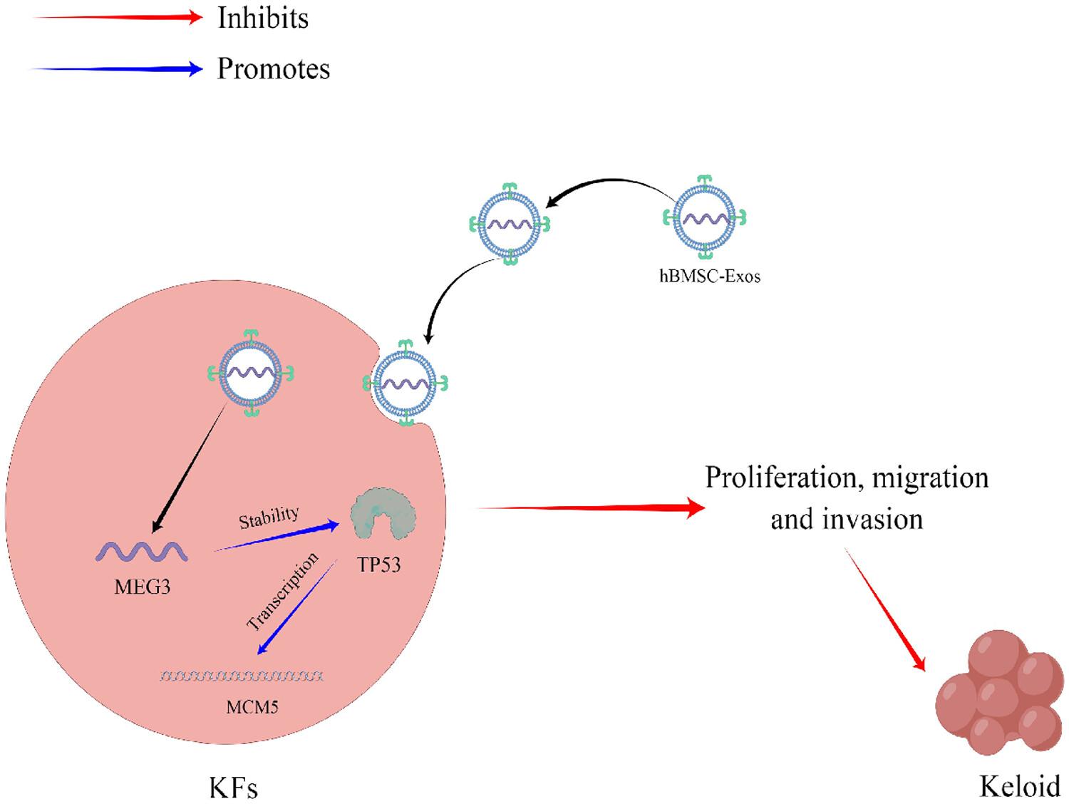
Exosomes were isolated from hBMSC, and keloid fibroblasts (KFs) were treated with hBMSC-Exos. Cell counting kit-8, wound healing, transwell invasion, immunofluorescence, and western blot assays were conducted to study the malignant phenotype of KFs. Mice were induced with keloids and treated with hBMSC-Exos. The effect of hBMSC-Exos on keloid formation in vivo was evaluated by hematoxylin and eosin staining, Masson staining, immunohistochemistry, and western blotting. The GSE182192 dataset was screened for differentially expressed long non-coding RNA during keloid formation. Next, maternally expressed gene 3 (MEG3) was knocked down in hBMSC to obtain hBMSC-Exossh-MEG3. The molecular mechanism of MEG3 was investigated by bioinformatic screening, and the relationship between MEG3 and TP53 or MCM5 was verified.
Results
hBMSC-Exos inhibited the malignant proliferation, migration, and invasion of KFs at same time as promoting their apoptosis, Moreover, hBMSC-Exos reduced the expression of fibrosis- and collagen-related proteins in the cells and the formation of keloids caused by KFs. The reduction in MEG3 enrichment in hBMSC-Exos weakened the inhibitory effect of hBMSC-Exos on KF activity. hBMSC-Exos delivered MEG3 to promote MCM5 transcription by TP53 in KFs. Overexpression of MCM5 in KFs reversed the effects of hBMSC-Exossh-MEG3, leading to reduced KF activity.
Conclusions
hBMSC-Exos delivered MEG3 to promote the protein stability of TP53, thereby activating MCM5 and promoting KF activity.
期刊介绍:
The aims and scope of The Journal of Gene Medicine include cutting-edge science of gene transfer and its applications in gene and cell therapy, genome editing with precision nucleases, epigenetic modifications of host genome by small molecules, siRNA, microRNA and other noncoding RNAs as therapeutic gene-modulating agents or targets, biomarkers for precision medicine, and gene-based prognostic/diagnostic studies.
Key areas of interest are the design of novel synthetic and viral vectors, novel therapeutic nucleic acids such as mRNA, modified microRNAs and siRNAs, antagomirs, aptamers, antisense and exon-skipping agents, refined genome editing tools using nucleic acid /protein combinations, physically or biologically targeted delivery and gene modulation, ex vivo or in vivo pharmacological studies including animal models, and human clinical trials.
Papers presenting research into the mechanisms underlying transfer and action of gene medicines, the application of the new technologies for stem cell modification or nucleic acid based vaccines, the identification of new genetic or epigenetic variations as biomarkers to direct precision medicine, and the preclinical/clinical development of gene/expression signatures indicative of diagnosis or predictive of prognosis are also encouraged.


 分享
分享
 求助内容:
求助内容: 应助结果提醒方式:
 扫码关注我们
