Hai Xin*, Ben M. Ferguson, Boyang Wan, D S Abdullah Al Maruf, William T. Lewin, Kai Cheng, Hedi V. Kruse, David Leinkram, Krishnan Parthasarathi, Innes K. Wise, Catriona Froggatt, Jeremy M. Crook, David R. McKenzie, Qing Li and Jonathan R. Clark,
{"title":"A Preclinical Trial Protocol Using an Ovine Model to Assess Scaffold Implant Biomaterials for Repair of Critical-Sized Mandibular Defects","authors":"Hai Xin*, Ben M. Ferguson, Boyang Wan, D S Abdullah Al Maruf, William T. Lewin, Kai Cheng, Hedi V. Kruse, David Leinkram, Krishnan Parthasarathi, Innes K. Wise, Catriona Froggatt, Jeremy M. Crook, David R. McKenzie, Qing Li and Jonathan R. Clark, ","doi":"10.1021/acsbiomaterials.4c00262","DOIUrl":null,"url":null,"abstract":"<p >The present work describes a preclinical trial (<i>in silico</i>, <i>in vivo</i> and <i>in vitro</i>) protocol to assess the biomechanical performance and osteogenic capability of 3D-printed polymeric scaffolds implants used to repair partial defects in a sheep mandible. The protocol spans multiple steps of the medical device development pipeline, including initial concept design of the scaffold implant, digital twin <i>in silico</i> finite element modeling, manufacturing of the device prototype, <i>in vivo</i> device implantation, and <i>in vitro</i> laboratory mechanical testing. First, a patient-specific one-body scaffold implant used for reconstructing a critical-sized defect along the lower border of the sheep mandible ramus was designed using on computed-tomographic (CT) imagery and computer-aided design software. Next, the biomechanical performance of the implant was predicted numerically by simulating physiological load conditions in a digital twin <i>in silico</i> finite element model of the sheep mandible. This allowed for possible redesigning of the implant prior to commencing <i>in vivo</i> experimentation. Then, two types of polymeric biomaterials were used to manufacture the mandibular scaffold implants: poly ether ether ketone (PEEK) and poly ether ketone (PEK) printed with fused deposition modeling (FDM) and selective laser sintering (SLS), respectively. Then, after being implanted for 13 weeks <i>in</i> vivo, the implant and surrounding bone tissue was harvested and microCT scanned to visualize and quantify neo-tissue formation in the porous space of the scaffold. Finally, the implant and local bone tissue was assessed by <i>in vitro</i> laboratory mechanical testing to quantify the osteointegration. The protocol consists of six component procedures: (i) scaffold design and finite element analysis to predict its biomechanical response, (ii) scaffold fabrication with FDM and SLS 3D printing, (iii) surface treatment of the scaffold with plasma immersion ion implantation (PIII) techniques, (iv) ovine mandibular implantation, (v) postoperative sheep recovery, euthanasia, and harvesting of the scaffold and surrounding host bone, microCT scanning, and (vi) <i>in vitro</i> laboratory mechanical tests of the harvested scaffolds. The results of microCT imagery and 3-point mechanical bend testing demonstrate that PIII-SLS-PEK is a promising biomaterial for the manufacturing of scaffold implants to enhance the bone-scaffold contact and bone ingrowth in porous scaffold implants. MicroCT images of the harvested implant and surrounding bone tissue showed encouraging new bone growth at the scaffold-bone interface and inside the porous network of the lattice structure of the SLS-PEK scaffolds.</p>","PeriodicalId":8,"journal":{"name":"ACS Biomaterials Science & Engineering","volume":"10 5","pages":"2863–2879"},"PeriodicalIF":5.5000,"publicationDate":"2024-05-02","publicationTypes":"Journal Article","fieldsOfStudy":null,"isOpenAccess":false,"openAccessPdf":"","citationCount":"0","resultStr":null,"platform":"Semanticscholar","paperid":null,"PeriodicalName":"ACS Biomaterials Science & Engineering","FirstCategoryId":"5","ListUrlMain":"https://pubs.acs.org/doi/10.1021/acsbiomaterials.4c00262","RegionNum":2,"RegionCategory":"医学","ArticlePicture":[],"TitleCN":null,"AbstractTextCN":null,"PMCID":null,"EPubDate":"","PubModel":"","JCR":"Q2","JCRName":"MATERIALS SCIENCE, BIOMATERIALS","Score":null,"Total":0}
引用次数: 0
Abstract
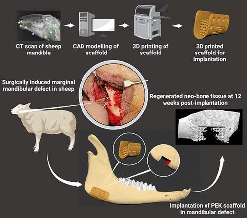
The present work describes a preclinical trial (in silico, in vivo and in vitro) protocol to assess the biomechanical performance and osteogenic capability of 3D-printed polymeric scaffolds implants used to repair partial defects in a sheep mandible. The protocol spans multiple steps of the medical device development pipeline, including initial concept design of the scaffold implant, digital twin in silico finite element modeling, manufacturing of the device prototype, in vivo device implantation, and in vitro laboratory mechanical testing. First, a patient-specific one-body scaffold implant used for reconstructing a critical-sized defect along the lower border of the sheep mandible ramus was designed using on computed-tomographic (CT) imagery and computer-aided design software. Next, the biomechanical performance of the implant was predicted numerically by simulating physiological load conditions in a digital twin in silico finite element model of the sheep mandible. This allowed for possible redesigning of the implant prior to commencing in vivo experimentation. Then, two types of polymeric biomaterials were used to manufacture the mandibular scaffold implants: poly ether ether ketone (PEEK) and poly ether ketone (PEK) printed with fused deposition modeling (FDM) and selective laser sintering (SLS), respectively. Then, after being implanted for 13 weeks in vivo, the implant and surrounding bone tissue was harvested and microCT scanned to visualize and quantify neo-tissue formation in the porous space of the scaffold. Finally, the implant and local bone tissue was assessed by in vitro laboratory mechanical testing to quantify the osteointegration. The protocol consists of six component procedures: (i) scaffold design and finite element analysis to predict its biomechanical response, (ii) scaffold fabrication with FDM and SLS 3D printing, (iii) surface treatment of the scaffold with plasma immersion ion implantation (PIII) techniques, (iv) ovine mandibular implantation, (v) postoperative sheep recovery, euthanasia, and harvesting of the scaffold and surrounding host bone, microCT scanning, and (vi) in vitro laboratory mechanical tests of the harvested scaffolds. The results of microCT imagery and 3-point mechanical bend testing demonstrate that PIII-SLS-PEK is a promising biomaterial for the manufacturing of scaffold implants to enhance the bone-scaffold contact and bone ingrowth in porous scaffold implants. MicroCT images of the harvested implant and surrounding bone tissue showed encouraging new bone growth at the scaffold-bone interface and inside the porous network of the lattice structure of the SLS-PEK scaffolds.
期刊介绍:
ACS Biomaterials Science & Engineering is the leading journal in the field of biomaterials, serving as an international forum for publishing cutting-edge research and innovative ideas on a broad range of topics:
Applications and Health – implantable tissues and devices, prosthesis, health risks, toxicology
Bio-interactions and Bio-compatibility – material-biology interactions, chemical/morphological/structural communication, mechanobiology, signaling and biological responses, immuno-engineering, calcification, coatings, corrosion and degradation of biomaterials and devices, biophysical regulation of cell functions
Characterization, Synthesis, and Modification – new biomaterials, bioinspired and biomimetic approaches to biomaterials, exploiting structural hierarchy and architectural control, combinatorial strategies for biomaterials discovery, genetic biomaterials design, synthetic biology, new composite systems, bionics, polymer synthesis
Controlled Release and Delivery Systems – biomaterial-based drug and gene delivery, bio-responsive delivery of regulatory molecules, pharmaceutical engineering
Healthcare Advances – clinical translation, regulatory issues, patient safety, emerging trends
Imaging and Diagnostics – imaging agents and probes, theranostics, biosensors, monitoring
Manufacturing and Technology – 3D printing, inks, organ-on-a-chip, bioreactor/perfusion systems, microdevices, BioMEMS, optics and electronics interfaces with biomaterials, systems integration
Modeling and Informatics Tools – scaling methods to guide biomaterial design, predictive algorithms for structure-function, biomechanics, integrating bioinformatics with biomaterials discovery, metabolomics in the context of biomaterials
Tissue Engineering and Regenerative Medicine – basic and applied studies, cell therapies, scaffolds, vascularization, bioartificial organs, transplantation and functionality, cellular agriculture


 分享
分享
 求助内容:
求助内容: 应助结果提醒方式:
 扫码关注我们
