Mohammad Sadegh Masoudi, Sina Zoghi, Reza Taheri, Adrina Habibzadeh, Ali Ansari
{"title":"A Novel Skin Incision for Posterior Fossa Midline and Paramedian Lesions: A Technical Note and Case Series.","authors":"Mohammad Sadegh Masoudi, Sina Zoghi, Reza Taheri, Adrina Habibzadeh, Ali Ansari","doi":"10.30476/BEAT.2023.100324.1465","DOIUrl":null,"url":null,"abstract":"<p><p>Approaching posterior fossa pathologies is fairly challenging. Poor exposure, cerebrospinal fluid (CSF) leak following surgery, post-operative suboccipital and neck pain, as well as wound healing are all common complications following traditional suboccipital midline incision. Herein, we present a novel incision for approaching posterior fossa pathologies. The incision is shaped like a question mark and makes a musculofascial flap supplied by the occipital artery on top as well as a wide area for craniotomy. In our technique, the dura is also incised in a question mark-shaped manner. The new incision was used to operate on three patients who had masses in the posterior fossa. Following surgeries, none of the patients experienced any adverse events such as CSF leak, wound complications, severe suboccipital pain, and neck instability. This new incision not only facilitates approaching pathologies in the posterior fossa by providing wider exposure but also enables us to perform watertight dural closure, which reduces CSF leak. Furthermore, as the muscular incision provides a sufficient area for craniotomy, muscular retraction can be minimized to reduce post-operative pain. Moreover, unlike the midline avascular incision, the flap is well supplied by the occipital artery, which facilitates the healing procedure.</p>","PeriodicalId":9333,"journal":{"name":"Bulletin of emergency and trauma","volume":"12 1","pages":"42-45"},"PeriodicalIF":0.0000,"publicationDate":"2024-01-01","publicationTypes":"Journal Article","fieldsOfStudy":null,"isOpenAccess":false,"openAccessPdf":"https://www.ncbi.nlm.nih.gov/pmc/articles/PMC11057448/pdf/","citationCount":"0","resultStr":null,"platform":"Semanticscholar","paperid":null,"PeriodicalName":"Bulletin of emergency and trauma","FirstCategoryId":"1085","ListUrlMain":"https://doi.org/10.30476/BEAT.2023.100324.1465","RegionNum":0,"RegionCategory":null,"ArticlePicture":[],"TitleCN":null,"AbstractTextCN":null,"PMCID":null,"EPubDate":"","PubModel":"","JCR":"","JCRName":"","Score":null,"Total":0}
引用次数: 0
Abstract
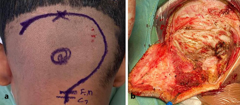
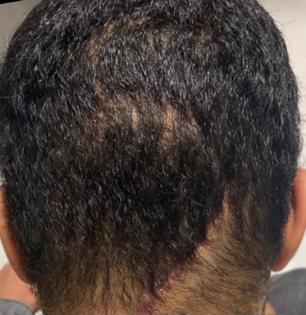
Approaching posterior fossa pathologies is fairly challenging. Poor exposure, cerebrospinal fluid (CSF) leak following surgery, post-operative suboccipital and neck pain, as well as wound healing are all common complications following traditional suboccipital midline incision. Herein, we present a novel incision for approaching posterior fossa pathologies. The incision is shaped like a question mark and makes a musculofascial flap supplied by the occipital artery on top as well as a wide area for craniotomy. In our technique, the dura is also incised in a question mark-shaped manner. The new incision was used to operate on three patients who had masses in the posterior fossa. Following surgeries, none of the patients experienced any adverse events such as CSF leak, wound complications, severe suboccipital pain, and neck instability. This new incision not only facilitates approaching pathologies in the posterior fossa by providing wider exposure but also enables us to perform watertight dural closure, which reduces CSF leak. Furthermore, as the muscular incision provides a sufficient area for craniotomy, muscular retraction can be minimized to reduce post-operative pain. Moreover, unlike the midline avascular incision, the flap is well supplied by the occipital artery, which facilitates the healing procedure.
期刊介绍:
BEAT: Bulletin of Emergency And Trauma is an international, peer-reviewed, quarterly journal coping with original research contributing to the field of emergency medicine and trauma. BEAT is the official journal of the Trauma Research Center (TRC) of Shiraz University of Medical Sciences (SUMS), Hungarian Trauma Society (HTS) and Lusitanian Association for Trauma and Emergency Surgery (ALTEC/LATES) aiming to be a publication of international repute that serves as a medium for dissemination and exchange of scientific knowledge in the emergency medicine and trauma. The aim of BEAT is to publish original research focusing on practicing and training of emergency medicine and trauma to publish peer-reviewed articles of current international interest in the form of original articles, brief communications, reviews, case reports, clinical images, and letters.


 分享
分享

 求助内容:
求助内容: 应助结果提醒方式:
 扫码关注我们
