Nivedita Sarveswaran, Yunisa Pamela, Akhila A N Reddy, Akash P Mustari, Anchala Parthasarathi, Anthony J Mancini, Anuradha Bishnoi, Arun C Inamadar, Bayanne Olabi, Fiona Browne, Gargi N Deshmukh, Kenneth McWilliam, Keshavamurthy Vinay, Sahana Srinivas, Samantha Ibbs, Sivakumar Natarajan, Vadlamudi R Rao, Vijay Zawar, Vykuntaraju K Gowda, Samiha S Shaikh, Celia Moss, Christopher G Woods, Ichrak Drissi
{"title":"Midfacial toddler excoriation syndrome (MiTES): case series, diagnostic criteria and evidence for a pathogenic mechanism.","authors":"Nivedita Sarveswaran, Yunisa Pamela, Akhila A N Reddy, Akash P Mustari, Anchala Parthasarathi, Anthony J Mancini, Anuradha Bishnoi, Arun C Inamadar, Bayanne Olabi, Fiona Browne, Gargi N Deshmukh, Kenneth McWilliam, Keshavamurthy Vinay, Sahana Srinivas, Samantha Ibbs, Sivakumar Natarajan, Vadlamudi R Rao, Vijay Zawar, Vykuntaraju K Gowda, Samiha S Shaikh, Celia Moss, Christopher G Woods, Ichrak Drissi","doi":"10.1093/bjd/ljae151","DOIUrl":null,"url":null,"abstract":"<p><strong>Background: </strong>PRDM12 polyalanine tract expansions cause two different disorders: midfacial toddler excoriation syndrome (MiTES; itch with normal pain sensation associated with 18 homozygous alanines (18A); and congenital insensitivity to pain (CIP) with normal itch associated with 19 homozygous alanines (19A). Knowledge of the phenotype, genotype and disease mechanism of MiTES is incomplete. Why 18A vs. 19A PRDM12 can cause almost opposite phenotypes is unknown; no other polyalanine or polyglutamine tract expansion disease causes two such disparate phenotypes.</p><p><strong>Objectives: </strong>To assess the genotype and phenotype of nine new, nine atypical and six previously reported patients diagnosed with MiTES.</p><p><strong>Methods: </strong>Using cell lines with homozygous PR domain zinc finger protein 12 (PRDM12) containing 12 alanines (12A; normal), 18A (MiTES) and 19A (CIP), we examined PRDM12 aggregation and subcellular localization by image-separation confocal microscopy and subcellular fractionation Western blotting.</p><p><strong>Results: </strong>MiTES presents in the first year of life; in all cases the condition regresses over the first decade, leaving scarring. The MiTES phenotype is highly distinctive. Features overlapping with PRDM12 CIP are rarely found. The genotype-phenotype study of the PRDM12 polyalanine tract shows that having 7-15 alanines is normal; 16-18 alanines is associated with MiTES; 19 alanines leads to CIP; and no clinically atypical cases of MiTES had a polyalanine tract expansion. PRDM12 aggregation and subcellular localization differed significantly between 18A and normal 12A cell lines and between 18A and 19A cell lines. MiTES is a new protein-aggregation disease.</p><p><strong>Conclusions: </strong>We provide diagnostic criteria for MiTES and improved longitudinal data. MiTES and CIP are distinct phenotypes, despite their genotypes varying by a single alanine in the PRDM12 polyalanine tract. We found clear distinctions between the cellular phenotypes of normal, MiTES and CIP cells. We hypothesize that the developmental environment of the trigeminal ganglion is unique and critically sensitive to pre- and postnatal levels of PRDM12.</p>","PeriodicalId":9238,"journal":{"name":"British Journal of Dermatology","volume":" ","pages":"437-446"},"PeriodicalIF":9.6000,"publicationDate":"2024-08-14","publicationTypes":"Journal Article","fieldsOfStudy":null,"isOpenAccess":false,"openAccessPdf":"https://www.ncbi.nlm.nih.gov/pmc/articles/PMC11324070/pdf/","citationCount":"0","resultStr":null,"platform":"Semanticscholar","paperid":null,"PeriodicalName":"British Journal of Dermatology","FirstCategoryId":"3","ListUrlMain":"https://doi.org/10.1093/bjd/ljae151","RegionNum":1,"RegionCategory":"医学","ArticlePicture":[],"TitleCN":null,"AbstractTextCN":null,"PMCID":null,"EPubDate":"","PubModel":"","JCR":"Q1","JCRName":"DERMATOLOGY","Score":null,"Total":0}
引用次数: 0
Abstract
Background: PRDM12 polyalanine tract expansions cause two different disorders: midfacial toddler excoriation syndrome (MiTES; itch with normal pain sensation associated with 18 homozygous alanines (18A); and congenital insensitivity to pain (CIP) with normal itch associated with 19 homozygous alanines (19A). Knowledge of the phenotype, genotype and disease mechanism of MiTES is incomplete. Why 18A vs. 19A PRDM12 can cause almost opposite phenotypes is unknown; no other polyalanine or polyglutamine tract expansion disease causes two such disparate phenotypes.
Objectives: To assess the genotype and phenotype of nine new, nine atypical and six previously reported patients diagnosed with MiTES.
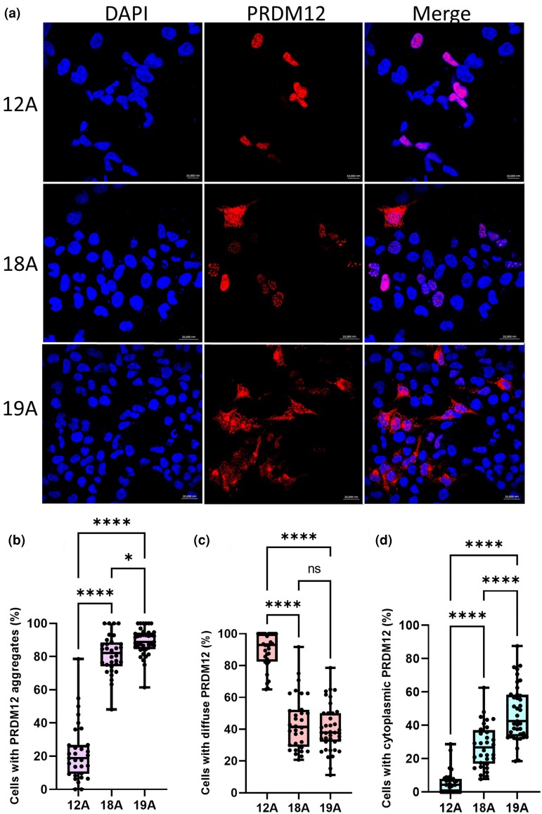
Methods: Using cell lines with homozygous PR domain zinc finger protein 12 (PRDM12) containing 12 alanines (12A; normal), 18A (MiTES) and 19A (CIP), we examined PRDM12 aggregation and subcellular localization by image-separation confocal microscopy and subcellular fractionation Western blotting.
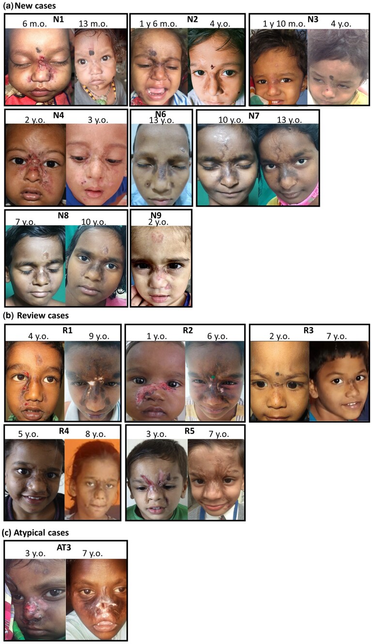
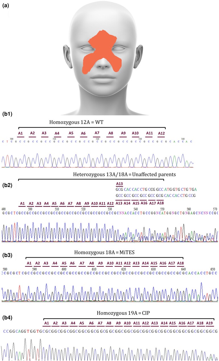
Results: MiTES presents in the first year of life; in all cases the condition regresses over the first decade, leaving scarring. The MiTES phenotype is highly distinctive. Features overlapping with PRDM12 CIP are rarely found. The genotype-phenotype study of the PRDM12 polyalanine tract shows that having 7-15 alanines is normal; 16-18 alanines is associated with MiTES; 19 alanines leads to CIP; and no clinically atypical cases of MiTES had a polyalanine tract expansion. PRDM12 aggregation and subcellular localization differed significantly between 18A and normal 12A cell lines and between 18A and 19A cell lines. MiTES is a new protein-aggregation disease.
Conclusions: We provide diagnostic criteria for MiTES and improved longitudinal data. MiTES and CIP are distinct phenotypes, despite their genotypes varying by a single alanine in the PRDM12 polyalanine tract. We found clear distinctions between the cellular phenotypes of normal, MiTES and CIP cells. We hypothesize that the developmental environment of the trigeminal ganglion is unique and critically sensitive to pre- and postnatal levels of PRDM12.
期刊介绍:
The British Journal of Dermatology (BJD) is committed to publishing the highest quality dermatological research. Through its publications, the journal seeks to advance the understanding, management, and treatment of skin diseases, ultimately aiming to improve patient outcomes.


 分享
分享
 求助内容:
求助内容: 应助结果提醒方式:
 扫码关注我们
