Radiographic Detection of Post-Traumatic Bone Fractures: Contribution of Artificial Intelligence Software to the Analysis of Senior and Junior Radiologists.
Andrea Dell'Aria, Denis Tack, Najat Saddiki, Sonia Makdoud, Jean Alexiou, François-Xavier De Hemptinne, Ivan Berkenbaum, Carine Neugroschl, Nunzia Tacelli
{"title":"Radiographic Detection of Post-Traumatic Bone Fractures: Contribution of Artificial Intelligence Software to the Analysis of Senior and Junior Radiologists.","authors":"Andrea Dell'Aria, Denis Tack, Najat Saddiki, Sonia Makdoud, Jean Alexiou, François-Xavier De Hemptinne, Ivan Berkenbaum, Carine Neugroschl, Nunzia Tacelli","doi":"10.5334/jbsr.3574","DOIUrl":null,"url":null,"abstract":"<p><strong>Objectives: </strong>The aims of this study were: (a) to evaluate the performance of an artificial intelligence (AI) software package (Boneview Trauma, Gleamer) for the detection of post-traumatic bone fractures in radiography as a standalone; (b) used by two radiologists (osteoarticular senior and junior); and (c) to determine to whom AI would be most helpful.</p><p><strong>Materials and methods: </strong>Within 14 days of a trauma, 101 consecutive patients underwent radiographic examination of the upper or lower limbs. The definite diagnosis for identifying fractures was: (a) radio-clinical consensus between the radiologist on-call who analyzed the images and the orthopedist (Group 1); (b) Cone Beam computed tomography (CBCT) exploration of the area of interest, in case of doubts or absence of consensus (Group 2). Independently of this diagnosis for both groups, the radiographic images were separately analyzed by two radiologists (osteoarticular senior: SR; junior: JR) prior without, and thereafter with the results of AI.</p><p><strong>Results: </strong>AI performed better than the radiologists in detecting common fractures (Group 1), but not subtle fractures (Group 2). In association with AI, both radiologists increased their overall performances in both groups, whereas this increase was significantly higher for the JR (<i>p</i> < 0.05).</p><p><strong>Conclusion: </strong>AI is reliable for common radiographic fracture identification and is a useful learning tool for radiologists in training. However, the software's overall performance does not exceed that of an osteoarticular senior radiologist, particularly in case of subtle lesions.</p>","PeriodicalId":55987,"journal":{"name":"Journal of the Belgian Society of Radiology","volume":"108 1","pages":"44"},"PeriodicalIF":1.3000,"publicationDate":"2024-04-25","publicationTypes":"Journal Article","fieldsOfStudy":null,"isOpenAccess":false,"openAccessPdf":"https://www.ncbi.nlm.nih.gov/pmc/articles/PMC11049681/pdf/","citationCount":"0","resultStr":null,"platform":"Semanticscholar","paperid":null,"PeriodicalName":"Journal of the Belgian Society of Radiology","FirstCategoryId":"3","ListUrlMain":"https://doi.org/10.5334/jbsr.3574","RegionNum":4,"RegionCategory":"医学","ArticlePicture":[],"TitleCN":null,"AbstractTextCN":null,"PMCID":null,"EPubDate":"2024/1/1 0:00:00","PubModel":"eCollection","JCR":"Q4","JCRName":"RADIOLOGY, NUCLEAR MEDICINE & MEDICAL IMAGING","Score":null,"Total":0}
引用次数: 0
Abstract
Objectives: The aims of this study were: (a) to evaluate the performance of an artificial intelligence (AI) software package (Boneview Trauma, Gleamer) for the detection of post-traumatic bone fractures in radiography as a standalone; (b) used by two radiologists (osteoarticular senior and junior); and (c) to determine to whom AI would be most helpful.
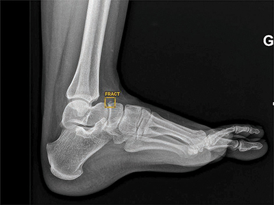
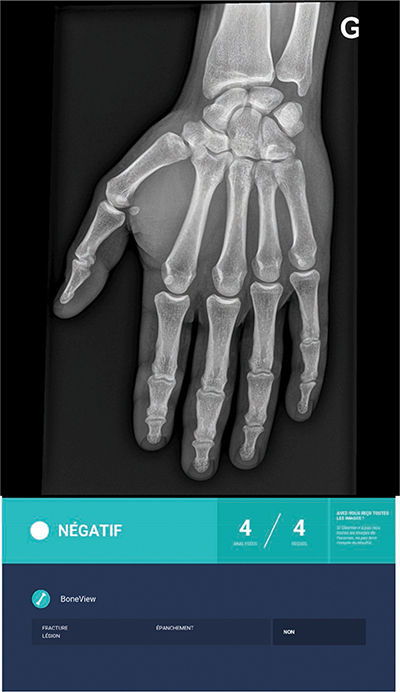
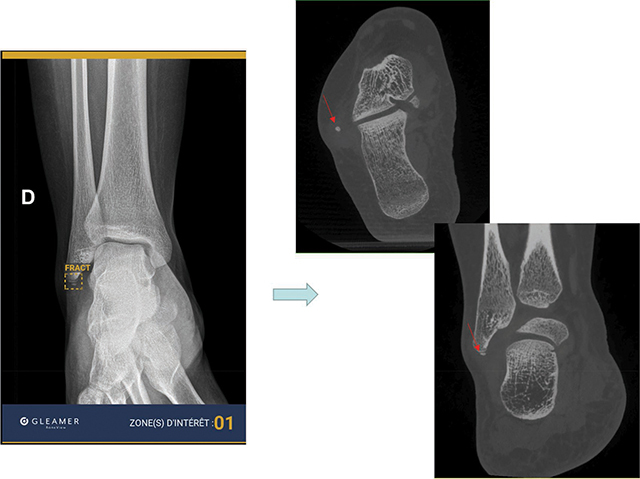
Materials and methods: Within 14 days of a trauma, 101 consecutive patients underwent radiographic examination of the upper or lower limbs. The definite diagnosis for identifying fractures was: (a) radio-clinical consensus between the radiologist on-call who analyzed the images and the orthopedist (Group 1); (b) Cone Beam computed tomography (CBCT) exploration of the area of interest, in case of doubts or absence of consensus (Group 2). Independently of this diagnosis for both groups, the radiographic images were separately analyzed by two radiologists (osteoarticular senior: SR; junior: JR) prior without, and thereafter with the results of AI.
Results: AI performed better than the radiologists in detecting common fractures (Group 1), but not subtle fractures (Group 2). In association with AI, both radiologists increased their overall performances in both groups, whereas this increase was significantly higher for the JR (p < 0.05).
Conclusion: AI is reliable for common radiographic fracture identification and is a useful learning tool for radiologists in training. However, the software's overall performance does not exceed that of an osteoarticular senior radiologist, particularly in case of subtle lesions.
期刊介绍:
The purpose of the Journal of the Belgian Society of Radiology is the publication of articles dealing with diagnostic and interventional radiology, related imaging techniques, allied sciences, and continuing education.


 分享
分享
 求助内容:
求助内容: 应助结果提醒方式:
 扫码关注我们
