Ming-Kai Hsieh, Chi-Yun Wang, Fu-Cheng Kao, Hui-Ting Su, Mei-Feng Chen, Tsung-Ting Tsai, Po-Liang Lai
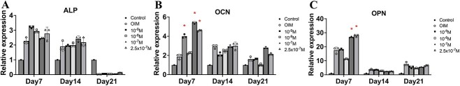
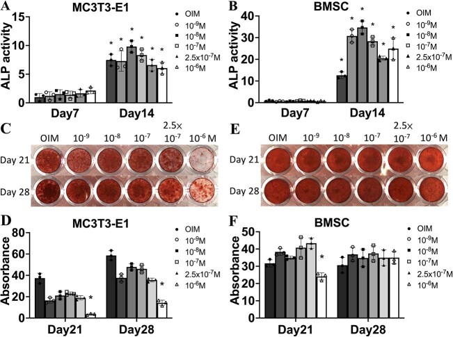
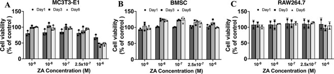
{"title":"Local application of zoledronate inhibits early bone resorption and promotes bone formation.","authors":"Ming-Kai Hsieh, Chi-Yun Wang, Fu-Cheng Kao, Hui-Ting Su, Mei-Feng Chen, Tsung-Ting Tsai, Po-Liang Lai","doi":"10.1093/jbmrpl/ziae031","DOIUrl":null,"url":null,"abstract":"<p><p>Nonunion resulting from early bone resorption is common after bone transplantation surgery. In these patients, instability or osteoporosis causes hyperactive catabolism relative to anabolism, leading to graft resorption instead of fusion. Systemic zoledronate administration inhibits osteoclastogenesis and is widely used to prevent osteoporosis; however, evidence on local zoledronate application is controversial due to osteoblast cytotoxicity, uncontrolled dosing regimens, and local release methods. We investigated the effects of zolendronate on osteoclastogenesis and osteogenesis and explored the corresponding signaling pathways. In vitro cytotoxicity and differentiation of MC3T3E1 cells, rat bone marrow stromal cells (BMSCs) and preosteoclasts (RAW264.7 cells) were evaluated with different zolendronate concentrations. In vivo bone regeneration ability was tested by transplanting different concentrations of zolendronate with β-tricalcium phosphate (TCP) bone substitute into rat femoral critical-sized bone defects. In vitro<i>,</i> zolendronate concentrations below 2.5 × 10<sup>-7</sup> M did not compromise viability in the three cell lines and did not promote osteogenic differentiation in MC3T3E1 cells and BMSCs. In RAW264.7 cells, zoledronate inhibited extracellular regulated protein kinases and c-Jun n-terminal kinase signaling, downregulating c-Fos and NFATc1 expression, with reduced expression of fusion-related dendritic cell‑specific transmembrane protein and osteoclast-specific Ctsk and tartrate-resistant acid phosphatase (. In vivo<i>,</i> histological staining revealed increased osteoid formation and neovascularization and reduced fibrotic tissue with 500 μM and 2000 μM zolendronate. More osteoclasts were found in the normal saline group after 6 weeks, and sequential osteoclast formation occurred after zoledronate treatment, indicating inhibition of bone resorption during early callus formation without inhibition of late-stage bone remodeling. In vivo<i>,</i> soaking β-TCP artificial bone with 500 μM or 2000 μM zoledronate is a promising approach for bone regeneration, with potential applications in bone transplantation.</p>","PeriodicalId":14611,"journal":{"name":"JBMR Plus","volume":"8 5","pages":"ziae031"},"PeriodicalIF":2.4000,"publicationDate":"2024-03-09","publicationTypes":"Journal Article","fieldsOfStudy":null,"isOpenAccess":false,"openAccessPdf":"https://www.ncbi.nlm.nih.gov/pmc/articles/PMC11008729/pdf/","citationCount":"0","resultStr":null,"platform":"Semanticscholar","paperid":null,"PeriodicalName":"JBMR Plus","FirstCategoryId":"1085","ListUrlMain":"https://doi.org/10.1093/jbmrpl/ziae031","RegionNum":0,"RegionCategory":null,"ArticlePicture":[],"TitleCN":null,"AbstractTextCN":null,"PMCID":null,"EPubDate":"2024/5/1 0:00:00","PubModel":"eCollection","JCR":"Q2","JCRName":"ENDOCRINOLOGY & METABOLISM","Score":null,"Total":0}
引用次数: 0
Abstract
Nonunion resulting from early bone resorption is common after bone transplantation surgery. In these patients, instability or osteoporosis causes hyperactive catabolism relative to anabolism, leading to graft resorption instead of fusion. Systemic zoledronate administration inhibits osteoclastogenesis and is widely used to prevent osteoporosis; however, evidence on local zoledronate application is controversial due to osteoblast cytotoxicity, uncontrolled dosing regimens, and local release methods. We investigated the effects of zolendronate on osteoclastogenesis and osteogenesis and explored the corresponding signaling pathways. In vitro cytotoxicity and differentiation of MC3T3E1 cells, rat bone marrow stromal cells (BMSCs) and preosteoclasts (RAW264.7 cells) were evaluated with different zolendronate concentrations. In vivo bone regeneration ability was tested by transplanting different concentrations of zolendronate with β-tricalcium phosphate (TCP) bone substitute into rat femoral critical-sized bone defects. In vitro, zolendronate concentrations below 2.5 × 10⁻⁷ M did not compromise viability in the three cell lines and did not promote osteogenic differentiation in MC3T3E1 cells and BMSCs. In RAW264.7 cells, zoledronate inhibited extracellular regulated protein kinases and c-Jun n-terminal kinase signaling, downregulating c-Fos and NFATc1 expression, with reduced expression of fusion-related dendritic cell‑specific transmembrane protein and osteoclast-specific Ctsk and tartrate-resistant acid phosphatase (. In vivo, histological staining revealed increased osteoid formation and neovascularization and reduced fibrotic tissue with 500 μM and 2000 μM zolendronate. More osteoclasts were found in the normal saline group after 6 weeks, and sequential osteoclast formation occurred after zoledronate treatment, indicating inhibition of bone resorption during early callus formation without inhibition of late-stage bone remodeling. In vivo, soaking β-TCP artificial bone with 500 μM or 2000 μM zoledronate is a promising approach for bone regeneration, with potential applications in bone transplantation.


 分享
分享
 求助内容:
求助内容: 应助结果提醒方式:
 扫码关注我们
