Huiru Li, Huawei Zhang, Li Yin, Feifei Zhang, Ziqi Chen, Taolin Chen, Zhiyun Jia, Qiyong Gong
{"title":"Altered cortical morphology in major depression disorder patients with suicidality.","authors":"Huiru Li, Huawei Zhang, Li Yin, Feifei Zhang, Ziqi Chen, Taolin Chen, Zhiyun Jia, Qiyong Gong","doi":"10.1093/psyrad/kkaa002","DOIUrl":null,"url":null,"abstract":"<p><strong>Background: </strong>Major depressive disorder (MDD) is associated with high risk of suicide, but the biological underpinnings of suicidality in MDD patients are far from conclusive. Previous neuroimaging studies using voxel-based morphometry (VBM) demonstrated that depressed individuals with suicidal thoughts or behaviors exhibit specific cortical structure alterations. To complement VBM findings, surface-based morphometry (SBM) can provide more details into gray matter structure, including the cortical complexity, cortical thickness and sulcal depth for brain images.</p><p><strong>Objective: </strong>This study aims to use SBM to investigate cortical morphology alterations to obtain evidence for neuroanatomical alterations in depressed patients with suicidality.</p><p><strong>Methods: </strong>Here, 3D T1-weighted MR images of brain from 39 healthy controls, 40 depressed patients without suicidality (patient controls), and 39 with suicidality (suicidal groups) were analyzed based on SBM to estimate the fractal dimension, gyrification index, sulcal depth, and cortical thickness using the Computational Anatomy Toolbox. Correlation analyses were performed between clinical data and cortical surface measurements from patients.</p><p><strong>Results: </strong>Surface-based morphometry showed decreased sulcal depth in the parietal, frontal, limbic, occipital and temporal regions and decreased fractal dimension in the frontal regions in depressed patients with suicidality compared to both healthy and patient controls. Additionally, in patients with depression, the sulcal depth of the left caudal anterior cingulate cortex was negatively correlated with Hamilton Depression Rating Scale scores.</p><p><strong>Conclusions: </strong>Depressed patients with suicidality had abnormal cortical morphology in some brain regions within the default mode network, frontolimbic circuitry and temporal regions. These structural deficits may be associated with the dysfunction of emotional processing and impulsivity control. This study provides insights into the underlying neurobiology of the suicidal brain.</p>","PeriodicalId":93496,"journal":{"name":"Psychoradiology","volume":"1 1","pages":"13-22"},"PeriodicalIF":2.9000,"publicationDate":"2021-03-18","publicationTypes":"Journal Article","fieldsOfStudy":null,"isOpenAccess":false,"openAccessPdf":"https://www.ncbi.nlm.nih.gov/pmc/articles/PMC10917214/pdf/","citationCount":"0","resultStr":null,"platform":"Semanticscholar","paperid":null,"PeriodicalName":"Psychoradiology","FirstCategoryId":"1085","ListUrlMain":"https://doi.org/10.1093/psyrad/kkaa002","RegionNum":0,"RegionCategory":null,"ArticlePicture":[],"TitleCN":null,"AbstractTextCN":null,"PMCID":null,"EPubDate":"2021/3/1 0:00:00","PubModel":"eCollection","JCR":"","JCRName":"","Score":null,"Total":0}
引用次数: 0
Abstract
Background: Major depressive disorder (MDD) is associated with high risk of suicide, but the biological underpinnings of suicidality in MDD patients are far from conclusive. Previous neuroimaging studies using voxel-based morphometry (VBM) demonstrated that depressed individuals with suicidal thoughts or behaviors exhibit specific cortical structure alterations. To complement VBM findings, surface-based morphometry (SBM) can provide more details into gray matter structure, including the cortical complexity, cortical thickness and sulcal depth for brain images.
Objective: This study aims to use SBM to investigate cortical morphology alterations to obtain evidence for neuroanatomical alterations in depressed patients with suicidality.
Methods: Here, 3D T1-weighted MR images of brain from 39 healthy controls, 40 depressed patients without suicidality (patient controls), and 39 with suicidality (suicidal groups) were analyzed based on SBM to estimate the fractal dimension, gyrification index, sulcal depth, and cortical thickness using the Computational Anatomy Toolbox. Correlation analyses were performed between clinical data and cortical surface measurements from patients.
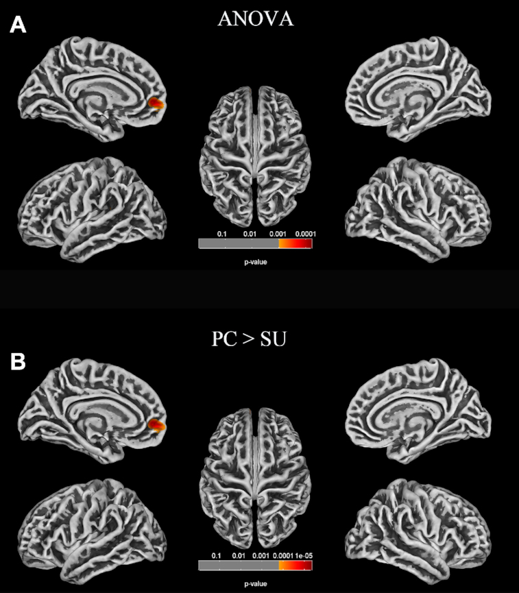
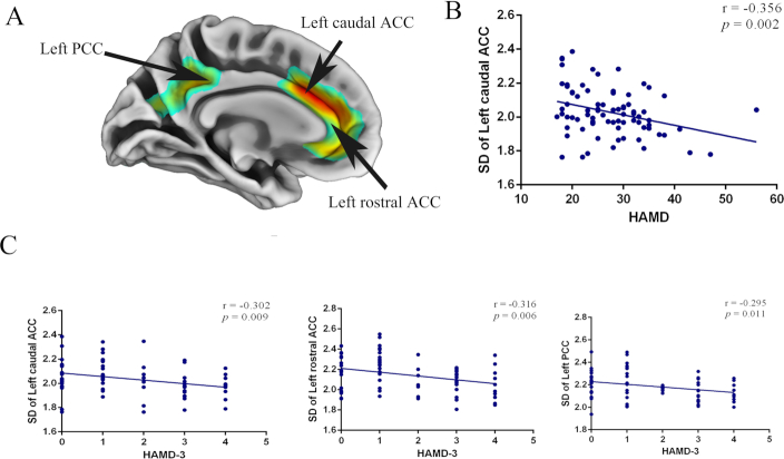
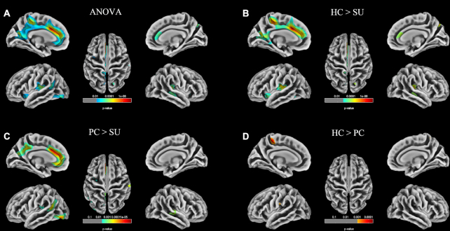
Results: Surface-based morphometry showed decreased sulcal depth in the parietal, frontal, limbic, occipital and temporal regions and decreased fractal dimension in the frontal regions in depressed patients with suicidality compared to both healthy and patient controls. Additionally, in patients with depression, the sulcal depth of the left caudal anterior cingulate cortex was negatively correlated with Hamilton Depression Rating Scale scores.
Conclusions: Depressed patients with suicidality had abnormal cortical morphology in some brain regions within the default mode network, frontolimbic circuitry and temporal regions. These structural deficits may be associated with the dysfunction of emotional processing and impulsivity control. This study provides insights into the underlying neurobiology of the suicidal brain.


 分享
分享

 求助内容:
求助内容: 应助结果提醒方式:
 扫码关注我们
