Scanning Electron Microscopy
Elizabeth R. Fischer, Bryan T. Hansen, Vinod Nair, Forrest H. Hoyt, Cindi L. Schwartz, David W. Dorward
{"title":"Scanning Electron Microscopy","authors":"Elizabeth R. Fischer, Bryan T. Hansen, Vinod Nair, Forrest H. Hoyt, Cindi L. Schwartz, David W. Dorward","doi":"10.1002/cpz1.1034","DOIUrl":null,"url":null,"abstract":"<p>Scanning electron microscopy (SEM) remains distinct in its ability to allow topographical visualization of structures. Key elements to consider for successful examination of biological specimens include appropriate preparative and imaging techniques. Chemical processing induces structural artifacts during specimen preparation, and several factors need to be considered when selecting fixation protocols to reduce these effects while retaining structures of interest. Particular care for proper dehydration of specimens is essential to minimize shrinkage and is necessary for placement under the high-vacuum environment required for routine operation of standard SEMs. Choice of substrate for mounting and coating specimens can reduce artifacts known as charging, and a basic understanding of microscope settings can optimize parameters to achieve desired results. This article describes fundamental techniques and tips for routine specimen preparation for a variety of biological specimens, preservation of labile or fragile structures, immune-labeling strategies, and microscope imaging parameters for optimal examination by SEM. © 2024 The Authors. Current Protocols published by Wiley Periodicals LLC.</p><p><b>Basic Protocol 1</b>: Chemical preparative techniques for preservation of biological specimens for examination by SEM</p><p><b>Alternate Protocol 1</b>: Practical considerations for the preparation of soft tissues</p><p><b>Alternate Protocol 2</b>: Removal of debris from the exoskeleton of invertebrates</p><p><b>Alternate Protocol 3</b>: Fixation of colonies grown on agar plates</p><p><b>Alternate Protocol 4</b>: Stabilization of polysaccharide structures with alcian blue and lysine</p><p><b>Alternate Protocol 5</b>: Preparation of non-adherent particulates in solution for SEM</p><p><b>Support Protocol 1</b>: Application of thin layer of adhesive on substrate to improve adherence</p><p><b>Support Protocol 2</b>: Poly-<span>L</span>-lysine coating specimen substrates for improved adherence</p><p><b>Support Protocol 3</b>: Microwave processing of biological specimens for examination by SEM</p><p><b>Basic Protocol 2</b>: Critical point drying of specimens</p><p><b>Alternate Protocol 6</b>: Chemical alternative to critical point drying</p><p><b>Basic Protocol 3</b>: Sputter coating</p><p><b>Alternate Protocol 7</b>: Improved bulk conductivity through “OTOTO”</p><p><b>Basic Protocol 4</b>: Immune-labeling strategies</p><p><b>Alternate Protocol 8</b>: Immune-labeling internal antigens with small gold probes</p><p><b>Alternate protocol 9</b>: Quantum dot or fluoronanogold preparations for correlative techniques</p><p><b>Basic Protocol 5</b>: Exposure of internal structures by mechanical fracturing</p><p><b>Basic Protocol 6</b>: Exposure of internal structures of tissues by fracturing with liquid nitrogen</p><p><b>Basic Protocol 7</b>: Anaglyph production from stereo pairs to produce 3D images</p>","PeriodicalId":93970,"journal":{"name":"Current protocols","volume":"4 5","pages":""},"PeriodicalIF":2.2000,"publicationDate":"2024-05-08","publicationTypes":"Journal Article","fieldsOfStudy":null,"isOpenAccess":false,"openAccessPdf":"https://onlinelibrary.wiley.com/doi/epdf/10.1002/cpz1.1034","citationCount":"0","resultStr":null,"platform":"Semanticscholar","paperid":null,"PeriodicalName":"Current protocols","FirstCategoryId":"1085","ListUrlMain":"https://currentprotocols.onlinelibrary.wiley.com/doi/10.1002/cpz1.1034","RegionNum":0,"RegionCategory":null,"ArticlePicture":[],"TitleCN":null,"AbstractTextCN":null,"PMCID":null,"EPubDate":"","PubModel":"","JCR":"","JCRName":"","Score":null,"Total":0}
引用次数: 0
Abstract
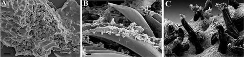
Scanning electron microscopy (SEM) remains distinct in its ability to allow topographical visualization of structures. Key elements to consider for successful examination of biological specimens include appropriate preparative and imaging techniques. Chemical processing induces structural artifacts during specimen preparation, and several factors need to be considered when selecting fixation protocols to reduce these effects while retaining structures of interest. Particular care for proper dehydration of specimens is essential to minimize shrinkage and is necessary for placement under the high-vacuum environment required for routine operation of standard SEMs. Choice of substrate for mounting and coating specimens can reduce artifacts known as charging, and a basic understanding of microscope settings can optimize parameters to achieve desired results. This article describes fundamental techniques and tips for routine specimen preparation for a variety of biological specimens, preservation of labile or fragile structures, immune-labeling strategies, and microscope imaging parameters for optimal examination by SEM. © 2024 The Authors. Current Protocols published by Wiley Periodicals LLC.
Basic Protocol 1: Chemical preparative techniques for preservation of biological specimens for examination by SEM
Alternate Protocol 1: Practical considerations for the preparation of soft tissues
Alternate Protocol 2: Removal of debris from the exoskeleton of invertebrates
Alternate Protocol 3: Fixation of colonies grown on agar plates
Alternate Protocol 4: Stabilization of polysaccharide structures with alcian blue and lysine
Alternate Protocol 5: Preparation of non-adherent particulates in solution for SEM
Support Protocol 1: Application of thin layer of adhesive on substrate to improve adherence
Support Protocol 2: Poly-L-lysine coating specimen substrates for improved adherence
Support Protocol 3: Microwave processing of biological specimens for examination by SEM
Basic Protocol 2: Critical point drying of specimens
Alternate Protocol 6: Chemical alternative to critical point drying
Basic Protocol 3: Sputter coating
Alternate Protocol 7: Improved bulk conductivity through “OTOTO”
Basic Protocol 4: Immune-labeling strategies
Alternate Protocol 8: Immune-labeling internal antigens with small gold probes
Alternate protocol 9: Quantum dot or fluoronanogold preparations for correlative techniques
Basic Protocol 5: Exposure of internal structures by mechanical fracturing
Basic Protocol 6: Exposure of internal structures of tissues by fracturing with liquid nitrogen
Basic Protocol 7: Anaglyph production from stereo pairs to produce 3D images

扫描电子显微镜
扫描电子显微镜(SEM)在对结构进行地形可视化方面的能力依然独树一帜。成功检查生物标本的关键因素包括适当的制备和成像技术。化学处理会在标本制备过程中产生结构伪影,因此在选择固定方案时需要考虑几个因素,以减少这些影响,同时保留感兴趣的结构。特别要注意标本的适当脱水,以尽量减少收缩,这对于将标本放置在标准扫描电子显微镜常规操作所需的高真空环境中非常必要。选择用于安装和涂布标本的基底可以减少被称为 "充电 "的伪影,而对显微镜设置的基本了解则可以优化参数,从而获得理想的结果。本文介绍了各种生物标本的常规标本制备、易变或易碎结构的保存、免疫标记策略和显微镜成像参数的基本技术和技巧,以便通过 SEM 进行最佳检查。© 2024 作者。当前协议》由 Wiley Periodicals LLC 出版。基本规程 1:用于保存生物标本以供扫描电镜检查的化学制备技术 备选规程 1:制备软组织的实用注意事项 备选规程 2:去除无脊椎动物外骨骼中的碎屑 备选规程 3:固定琼脂平板上生长的菌落 备选规程 4:稳定多糖结构备用方案 5:为扫描电子显微镜制备溶液中的非附着微粒 支持方案 1:在基底上涂抹薄层粘合剂以提高附着力 支持方案 2:在标本基底上涂抹聚 L-赖氨酸以提高附着力 支持方案 3:用微波处理生物标本以进行扫描电子显微镜分析 支持方案 4:用藻蓝和赖氨酸稳定多糖结构微波处理生物标本,供扫描电子显微镜检查 基本规程 2:标本的临界点干燥 替代规程 6:临界点干燥的化学替代方法 基本规程 3:溅射涂层 替代规程 7:通过 "OTOTO "提高批量导电性 基本规程 4:免疫标记策略 替代规程 8:免疫标记内部抗原替代方案 9:用于相关技术的量子点或氟化金制备 基本方案 5:通过机械压裂暴露内部结构 基本方案 6:通过液氮压裂暴露组织内部结构 基本方案 7:从立体图像对中生成 Anaglyph,以产生三维图像。
本文章由计算机程序翻译,如有差异,请以英文原文为准。


 分享
分享

 求助内容:
求助内容: 应助结果提醒方式:
 扫码关注我们
