{"title":"P3H4 Regulates Apoptosis and Autophagy of Breast Cancer Cells via BCL-2/BAX/Caspase-3 and AMPK/mTOR/ULK1 Signaling Pathways","authors":"T. Wang, M. M. Li, Z. Dong, D. M. Zhu","doi":"10.1134/s0026893324700237","DOIUrl":null,"url":null,"abstract":"<h3 data-test=\"abstract-sub-heading\">Abstract</h3><p>The role of prolyl 3-hydroxylase family member 4 (P3H4, or SC65) in breast cancer and the molecular mechanisms in which this protein was involved were investigated. For this purpose, microchips with cancerous and paracancerous tissues collected from 56 patients with breast cancer were constructed. The P3H4 protein expression was evaluated by immunohistochemistry. Cell lines with decreased and increased P3H4 expression were selected and divided into five groups: <i>P3H4</i> overexpression and corresponding negative control, <i>P3H4</i> knockout groups #1 and #2 and corresponding negative control. CCK8 assay, colony formation test, immunoblotting, scratch test, transwell test and flow cytometry were used to determine the relevant cell functions. P3H4 expression was higher in breast cancer cells than in paracancerous tissue. Compared with corresponding negative control, proliferative activity of the cells was inhibited at 24, 48 and 72 h, migration activity and invasion ability of the cells were reduced, autophagy and apoptosis in the cells were increased in <i>P3H4</i> knockout groups #1 and #2. <i>P3H4</i> knockout promoted apoptosis of breast cancer cells and inhibited their proliferation, migration, and invasion by activating the BCL-2/BAX/Caspase-3 and AMPK/mTOR pathways. <i>P3H4</i> knockout promoted apparently autophagy by activating the AMPK/mTOR/ULK1 pathway. However, <i>P3H4</i> overexpression could promote the proliferation, migration and invasion of breast cancer cells, and inhibited apoptosis and autophagy of mammary gland adenocarcinoma cells MDA-MB-231.</p>","PeriodicalId":18734,"journal":{"name":"Molecular Biology","volume":"109 1","pages":""},"PeriodicalIF":1.2000,"publicationDate":"2024-05-02","publicationTypes":"Journal Article","fieldsOfStudy":null,"isOpenAccess":false,"openAccessPdf":"","citationCount":"0","resultStr":null,"platform":"Semanticscholar","paperid":null,"PeriodicalName":"Molecular Biology","FirstCategoryId":"99","ListUrlMain":"https://doi.org/10.1134/s0026893324700237","RegionNum":4,"RegionCategory":"生物学","ArticlePicture":[],"TitleCN":null,"AbstractTextCN":null,"PMCID":null,"EPubDate":"","PubModel":"","JCR":"Q4","JCRName":"BIOCHEMISTRY & MOLECULAR BIOLOGY","Score":null,"Total":0}
引用次数: 0
Abstract
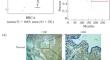
The role of prolyl 3-hydroxylase family member 4 (P3H4, or SC65) in breast cancer and the molecular mechanisms in which this protein was involved were investigated. For this purpose, microchips with cancerous and paracancerous tissues collected from 56 patients with breast cancer were constructed. The P3H4 protein expression was evaluated by immunohistochemistry. Cell lines with decreased and increased P3H4 expression were selected and divided into five groups: P3H4 overexpression and corresponding negative control, P3H4 knockout groups #1 and #2 and corresponding negative control. CCK8 assay, colony formation test, immunoblotting, scratch test, transwell test and flow cytometry were used to determine the relevant cell functions. P3H4 expression was higher in breast cancer cells than in paracancerous tissue. Compared with corresponding negative control, proliferative activity of the cells was inhibited at 24, 48 and 72 h, migration activity and invasion ability of the cells were reduced, autophagy and apoptosis in the cells were increased in P3H4 knockout groups #1 and #2. P3H4 knockout promoted apoptosis of breast cancer cells and inhibited their proliferation, migration, and invasion by activating the BCL-2/BAX/Caspase-3 and AMPK/mTOR pathways. P3H4 knockout promoted apparently autophagy by activating the AMPK/mTOR/ULK1 pathway. However, P3H4 overexpression could promote the proliferation, migration and invasion of breast cancer cells, and inhibited apoptosis and autophagy of mammary gland adenocarcinoma cells MDA-MB-231.
期刊介绍:
Molecular Biology is an international peer reviewed journal that covers a wide scope of problems in molecular, cell and computational biology including genomics, proteomics, bioinformatics, molecular virology and immunology, molecular development biology, molecular evolution and related areals. Molecular Biology publishes reviews, experimental and theoretical works. Every year, the journal publishes special issues devoted to most rapidly developing branches of physical-chemical biology and to the most outstanding scientists.


 分享
分享
 求助内容:
求助内容: 应助结果提醒方式:
 扫码关注我们
