Identify the benefits and caveats of combining minimal access approaches to the infratemporal fossa (ITF), such as the endoscopic transnasal, endoscopic transorbital, endoscopic transoral, and endoscopic sublabial transmaxillary approaches to address extensive lesions not amenable to a single approach. The study provides anatomical metrics including area of exposure and degree of surgical freedom.
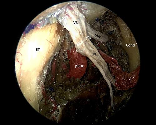
Five human cadaveric specimens (10 sides) were dissected to expose and methodically analyze the anatomical intricacies of the ITF using the following minimal access approaches: endoscopic transnasal transpterygoid (EETA), endoscopic sublabial transmaxillary, endoscopic transorbital via infraorbital foramen, and endoscopic transoral techniques. Area of exposure at the pterygopalatine fossa and surgical freedom at the ITF were obtained for each approach.
The endoscopic sublabial transmaxillary sinus and the combined approach afford a significantly greater exposure than an isolated EETA. The difference in exposure (mean) between the endoscopic sublabial transmaxillary and EETA was 1.62 ± 0.85 cm2 (p < 0.001), and the difference between the combined approach and EETA was 4.25 ± 0.85 cm2 (p < 0.001).
Combining minimal access endoscopic approaches to the ITF can provide significantly greater exposure than an isolated EETA; thus, providing enhanced access to address lesions with extensive involvement of the ITF, especially those with superolateral and inferolateral extensions. In addition, some approaches may have an adjunctive role to the resection, such as the endoscopic transoral approach offering the potential for early control of the internal maxillary artery and its branches, some of which may be supplying the tumor in the ITF; or the endoscopic transorbital approach yielding a direct line of sight to the superior ITF and middle cranial fossa.
NA.


