Sarmiza Elena Stanca , Selene Mogavero , Wolfgang Fritzsche , Christoph Krafft , Bernhard Hube , Jürgen Popp
{"title":"Isotope labeled 3D-Raman confocal imaging and atomic force microscopy study on epithelial cells interacting with the fungus Candida albicans","authors":"Sarmiza Elena Stanca , Selene Mogavero , Wolfgang Fritzsche , Christoph Krafft , Bernhard Hube , Jürgen Popp","doi":"10.1016/j.nano.2024.102750","DOIUrl":null,"url":null,"abstract":"<div><p>The human pathogenic fungus <em>Candida albicans</em> damages epithelial cells during superficial infections. Here we use three-dimensional-sequential-confocal Raman spectroscopic imaging and atomic force microscopy to investigate the interaction of <em>C. albicans</em> wild type cells, the secreted <em>C. albicans</em> peptide toxin candidalysin and mutant cells lacking candidalysin with epithelial cells. The candidalysin is responsible for epithelial cell damage and exhibits in its deuterated form an identifiable Raman signal in a frequency region distinct from the cellular frequency region. Vibration modes at 2100–2200 cm<sup>−1</sup> attributed to carbon‑deuterium bending and at 477 cm<sup>−1</sup>, attributed to the nitrogen‑deuterium out-of-plane bending, found around the nucleus, can be assigned to deuterated candidalysin. Atomic force microscopy visualized 100 nm deep lesions on the cell and force-distance curves indicate the higher adhesion on pore surrounding after incubation with candidalysin. Candidalysin targets the plasma membrane, but is also found inside of the cytosol of epithelial cells during <em>C. albicans</em> infection.</p></div>","PeriodicalId":19050,"journal":{"name":"Nanomedicine : nanotechnology, biology, and medicine","volume":"59 ","pages":"Article 102750"},"PeriodicalIF":4.6000,"publicationDate":"2024-05-09","publicationTypes":"Journal Article","fieldsOfStudy":null,"isOpenAccess":false,"openAccessPdf":"","citationCount":"0","resultStr":null,"platform":"Semanticscholar","paperid":null,"PeriodicalName":"Nanomedicine : nanotechnology, biology, and medicine","FirstCategoryId":"3","ListUrlMain":"https://www.sciencedirect.com/science/article/pii/S1549963424000194","RegionNum":2,"RegionCategory":"医学","ArticlePicture":[],"TitleCN":null,"AbstractTextCN":null,"PMCID":null,"EPubDate":"","PubModel":"","JCR":"Q2","JCRName":"MEDICINE, RESEARCH & EXPERIMENTAL","Score":null,"Total":0}
引用次数: 0
Abstract
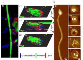
The human pathogenic fungus Candida albicans damages epithelial cells during superficial infections. Here we use three-dimensional-sequential-confocal Raman spectroscopic imaging and atomic force microscopy to investigate the interaction of C. albicans wild type cells, the secreted C. albicans peptide toxin candidalysin and mutant cells lacking candidalysin with epithelial cells. The candidalysin is responsible for epithelial cell damage and exhibits in its deuterated form an identifiable Raman signal in a frequency region distinct from the cellular frequency region. Vibration modes at 2100–2200 cm−1 attributed to carbon‑deuterium bending and at 477 cm−1, attributed to the nitrogen‑deuterium out-of-plane bending, found around the nucleus, can be assigned to deuterated candidalysin. Atomic force microscopy visualized 100 nm deep lesions on the cell and force-distance curves indicate the higher adhesion on pore surrounding after incubation with candidalysin. Candidalysin targets the plasma membrane, but is also found inside of the cytosol of epithelial cells during C. albicans infection.
期刊介绍:
The mission of Nanomedicine: Nanotechnology, Biology, and Medicine (Nanomedicine: NBM) is to promote the emerging interdisciplinary field of nanomedicine.
Nanomedicine: NBM is an international, peer-reviewed journal presenting novel, significant, and interdisciplinary theoretical and experimental results related to nanoscience and nanotechnology in the life and health sciences. Content includes basic, translational, and clinical research addressing diagnosis, treatment, monitoring, prediction, and prevention of diseases.


 分享
分享
 求助内容:
求助内容: 应助结果提醒方式:
 扫码关注我们
