Thomas Saliba, Marco Preziosi, Paolo Simoni, Alessandro De Leucio
{"title":"Fibromatosis Colli: A Thorough Description of Its MRI Characteristics and a Review of the Literature.","authors":"Thomas Saliba, Marco Preziosi, Paolo Simoni, Alessandro De Leucio","doi":"10.5334/jbsr.3270","DOIUrl":null,"url":null,"abstract":"<p><strong>Introduction: </strong>Fibromatosis colli (FC) is a rare pseudotumor of the sternocleidomastoid muscle with an incidence of 0.4%, generally diagnosed using ultrasound between 2 and 4 weeks of age. This is an important entity considering the clinical concerns it causes due to its appearance as a cervical mass with torticollis. Few magnetic resonance imaging (MRI) descriptions of its appearance have been made, with the existing reported cases being sporadic. We aim to provide a thorough description of this paediatric entity.</p><p><strong>Materials and methods: </strong>We conducted a retrospective study by searching our hospital's database for previous cases of FC where an MRI had been performed. We found six cases of FC where an MRI had been performed. Of these cases, five out of six were contrast-enhanced. We examined the MRIs to be able to discern and describe the MRI characteristics of FC.</p><p><strong>Results: </strong>We found that FC presents a T1 signal isointense to the muscle, a T2 signal hyperintense to the muscle, a variable diffusion signal and a thick enhancing peripheral ring after contrast administration.</p><p><strong>Discussion: </strong>Our results match what has been reported in the literature to date regarding the MRI signal of FC, confirming previous reports. However, we provide new data regarding the characteristic appearance post-enhancement, which was previously unreported.</p><p><strong>Conclusion: </strong>The MRI characteristics of FC have rarely been described, with only a few isolated case reports in the medical literature. We review the current literature, describe the key MRI characteristics of the pathology, and provide the most thorough description to date.</p>","PeriodicalId":55987,"journal":{"name":"Journal of the Belgian Society of Radiology","volume":"108 1","pages":"51"},"PeriodicalIF":1.3000,"publicationDate":"2024-05-07","publicationTypes":"Journal Article","fieldsOfStudy":null,"isOpenAccess":false,"openAccessPdf":"https://www.ncbi.nlm.nih.gov/pmc/articles/PMC11086591/pdf/","citationCount":"0","resultStr":null,"platform":"Semanticscholar","paperid":null,"PeriodicalName":"Journal of the Belgian Society of Radiology","FirstCategoryId":"3","ListUrlMain":"https://doi.org/10.5334/jbsr.3270","RegionNum":4,"RegionCategory":"医学","ArticlePicture":[],"TitleCN":null,"AbstractTextCN":null,"PMCID":null,"EPubDate":"2024/1/1 0:00:00","PubModel":"eCollection","JCR":"Q4","JCRName":"RADIOLOGY, NUCLEAR MEDICINE & MEDICAL IMAGING","Score":null,"Total":0}
引用次数: 0
Abstract
Introduction: Fibromatosis colli (FC) is a rare pseudotumor of the sternocleidomastoid muscle with an incidence of 0.4%, generally diagnosed using ultrasound between 2 and 4 weeks of age. This is an important entity considering the clinical concerns it causes due to its appearance as a cervical mass with torticollis. Few magnetic resonance imaging (MRI) descriptions of its appearance have been made, with the existing reported cases being sporadic. We aim to provide a thorough description of this paediatric entity.
Materials and methods: We conducted a retrospective study by searching our hospital's database for previous cases of FC where an MRI had been performed. We found six cases of FC where an MRI had been performed. Of these cases, five out of six were contrast-enhanced. We examined the MRIs to be able to discern and describe the MRI characteristics of FC.
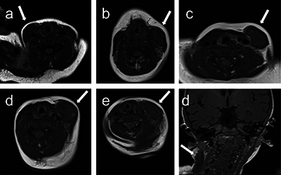
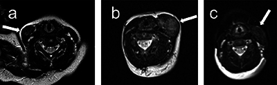
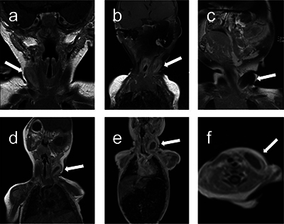
Results: We found that FC presents a T1 signal isointense to the muscle, a T2 signal hyperintense to the muscle, a variable diffusion signal and a thick enhancing peripheral ring after contrast administration.
Discussion: Our results match what has been reported in the literature to date regarding the MRI signal of FC, confirming previous reports. However, we provide new data regarding the characteristic appearance post-enhancement, which was previously unreported.
Conclusion: The MRI characteristics of FC have rarely been described, with only a few isolated case reports in the medical literature. We review the current literature, describe the key MRI characteristics of the pathology, and provide the most thorough description to date.
期刊介绍:
The purpose of the Journal of the Belgian Society of Radiology is the publication of articles dealing with diagnostic and interventional radiology, related imaging techniques, allied sciences, and continuing education.


 分享
分享
 求助内容:
求助内容: 应助结果提醒方式:
 扫码关注我们
