{"title":"Endoscopic Diagnosis of Epithelial Subtypes of Superficial Non-Ampullary Duodenal Epithelial Tumors using Magnifying Narrow-Band Imaging.","authors":"Yoshiyuki Kurata, Takashi Hirose, Naomi Kakushima, Masato Nakaguro, Yuki Okumura, Hitoshi Tanaka, Mitsuhiro Fujishiro, Hiroki Kawashima","doi":"10.1159/000539308","DOIUrl":null,"url":null,"abstract":"<p><strong>Introduction: </strong>Superficial non-ampullary duodenal epithelial tumors (SNADETs) include low-grade adenoma (LGA) and high-grade adenoma or carcinoma (HGA/Ca) and are classified into two different epithelial subtypes, gastric-type (G-type) and intestinal-type (I-type). We attempted to distinguish them by endoscopic characteristics including magnifying endoscopy with narrow-band imaging (M-NBI).</p><p><strong>Methods: </strong>Various endoscopic and M-NBI findings of 286 SNADETs were retrospectively reviewed and compared between G- and I-types and histological grades. M-NBI findings were divided into four patterns based on the following vascular patterns; absent, network, intrastructural vascular (ISV), and unclassified. Lesions displaying a single pattern were classified as mono-pattern and those displaying multiple patterns as mixed-pattern. Lesions showing CDX2 positivity were categorized as I-types and those showing MUC5AC or MUC6 positivity were categorized as G-types based on immunohistochemistry.</p><p><strong>Results: </strong>Among 286 lesions, 23 (8%) were G-type and 243 (85%) were I-type. More G-type lesions were located oral to papilla (91.3 vs. 45.6%, p < 0.001), and had protruding morphology compared to those of I-types (65.2 vs. 14.4%, p < 0.001). The major M-NBI pattern was ISV in G-type (78.2 vs. 26.3%, p < 0.001), and absent for I-type (0 vs. 34.5%, p = 0.003). Three endoscopic characteristics; location oral to papilla, protruding morphology, and major M-NBI pattern (ISV) were independent predictors for G-type. Mixed-pattern was more common in HGA/Ca than LGA for I-type (77.0 vs. 58.8%, p = 0.01); however, there was no difference for those in G-type.</p><p><strong>Conclusion: </strong>Endoscopic findings including M-NBI are useful to differentiate epithelial subtypes.</p>","PeriodicalId":11294,"journal":{"name":"Digestive Diseases","volume":" ","pages":"399-406"},"PeriodicalIF":2.1000,"publicationDate":"2024-01-01","publicationTypes":"Journal Article","fieldsOfStudy":null,"isOpenAccess":false,"openAccessPdf":"https://www.ncbi.nlm.nih.gov/pmc/articles/PMC11457973/pdf/","citationCount":"0","resultStr":null,"platform":"Semanticscholar","paperid":null,"PeriodicalName":"Digestive Diseases","FirstCategoryId":"3","ListUrlMain":"https://doi.org/10.1159/000539308","RegionNum":4,"RegionCategory":"医学","ArticlePicture":[],"TitleCN":null,"AbstractTextCN":null,"PMCID":null,"EPubDate":"2024/5/15 0:00:00","PubModel":"Epub","JCR":"Q3","JCRName":"GASTROENTEROLOGY & HEPATOLOGY","Score":null,"Total":0}
引用次数: 0
Abstract
Introduction: Superficial non-ampullary duodenal epithelial tumors (SNADETs) include low-grade adenoma (LGA) and high-grade adenoma or carcinoma (HGA/Ca) and are classified into two different epithelial subtypes, gastric-type (G-type) and intestinal-type (I-type). We attempted to distinguish them by endoscopic characteristics including magnifying endoscopy with narrow-band imaging (M-NBI).
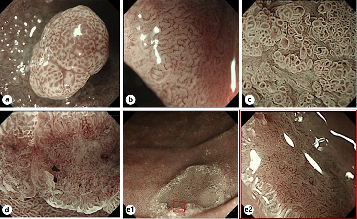
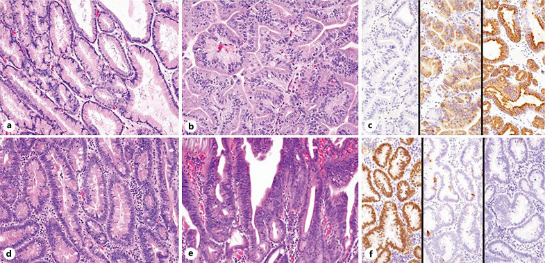
Methods: Various endoscopic and M-NBI findings of 286 SNADETs were retrospectively reviewed and compared between G- and I-types and histological grades. M-NBI findings were divided into four patterns based on the following vascular patterns; absent, network, intrastructural vascular (ISV), and unclassified. Lesions displaying a single pattern were classified as mono-pattern and those displaying multiple patterns as mixed-pattern. Lesions showing CDX2 positivity were categorized as I-types and those showing MUC5AC or MUC6 positivity were categorized as G-types based on immunohistochemistry.
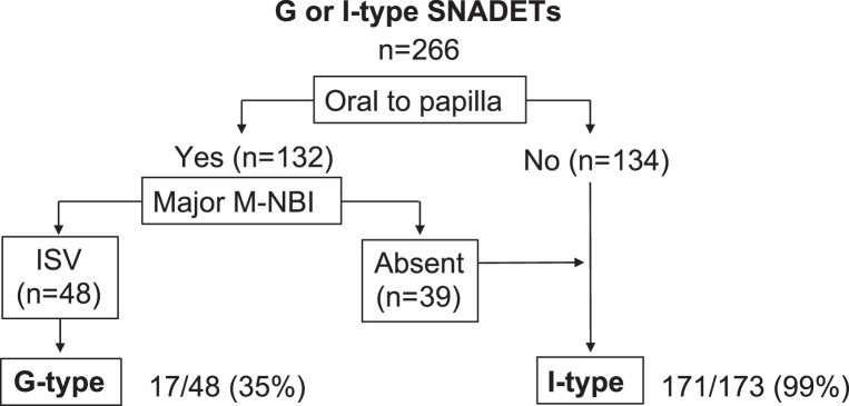
Results: Among 286 lesions, 23 (8%) were G-type and 243 (85%) were I-type. More G-type lesions were located oral to papilla (91.3 vs. 45.6%, p < 0.001), and had protruding morphology compared to those of I-types (65.2 vs. 14.4%, p < 0.001). The major M-NBI pattern was ISV in G-type (78.2 vs. 26.3%, p < 0.001), and absent for I-type (0 vs. 34.5%, p = 0.003). Three endoscopic characteristics; location oral to papilla, protruding morphology, and major M-NBI pattern (ISV) were independent predictors for G-type. Mixed-pattern was more common in HGA/Ca than LGA for I-type (77.0 vs. 58.8%, p = 0.01); however, there was no difference for those in G-type.
Conclusion: Endoscopic findings including M-NBI are useful to differentiate epithelial subtypes.
导言:浅表非髓质十二指肠上皮肿瘤(SNADETs)包括低级别腺瘤(LGA)和高级别腺瘤或癌(HGA/Ca),分为胃型(G型)和肠型(I型)两种不同的上皮亚型。我们试图通过内镜特征(包括放大内镜和窄带成像(M-NBI))来区分它们:方法:我们回顾性研究了286例SNADET的各种内镜和M-NBI结果,并对G型和I型以及组织学分级进行了比较。根据以下血管模式将 M-NBI 结果分为四种模式:无血管、网络状血管、结构内血管(ISV)和未分类。显示单一模式的病变被归类为单一模式,显示多种模式的病变被归类为混合模式。根据免疫组化结果,显示 CDX2 阳性的病变被归为 I 型,显示 MUC5AC 或 MUC6 阳性的病变被归为 G 型:在286个病灶中,23个(8%)为G型,243个(85%)为I型。与 I 型病变相比,更多的 G 型病变位于乳头口腔(91.3% vs 45.6%,P<0.001),且形态突出(65.2% vs 14.4%,P<0.001)。G 型的主要 M-NBI 模式是 ISV(78.2% vs 26.3%,P<0.001),而 I 型则没有(0% vs 34.5%,P=0.003)。三个内镜特征:乳头口腔位置、突出形态和主要 M-NBI 模式(ISV)是 G 型的独立预测因素。就 I 型而言,混合型在 HGA/Ca 比 LGA 中更为常见(77.0% vs 58.8%,P=0.01);但在 G 型中没有差异:结论:包括 M-NBI 在内的内镜检查结果有助于区分上皮亚型。
期刊介绍:
Each issue of this journal is dedicated to a special topic of current interest, covering both clinical and basic science topics in gastrointestinal function and disorders. The contents of each issue are comprehensive and reflect the state of the art, featuring editorials, reviews, mini reviews and original papers. These individual contributions encompass a variety of disciplines including all fields of gastroenterology. ''Digestive Diseases'' bridges the communication gap between advances made in the academic setting and their application in patient care. The journal is a valuable service for clinicians, specialists and physicians-in-training.


 分享
分享
 求助内容:
求助内容: 应助结果提醒方式:
 扫码关注我们
