{"title":"A simplified method of bacteriophage preparation for transmission electron microscope","authors":"Sepideh Meidaninikjeh , Parisa Mohammadi , Ameneh Elikaei","doi":"10.1016/j.jviromet.2024.114951","DOIUrl":null,"url":null,"abstract":"<div><p>Bacteriophages are viruses that infect bacteria. Researchers use different methods to study the characteristics of bacteriophages. Transmission electron microscope (TEM) is considered the best method to analyze these characteristics. However, the quality of TEM micrographs is significantly influenced by the preparation methods used to prepare the bacteriophages sample. In this study, researchers compared two different methods for preparing the bacteriophage samples. In one method was used SM buffer, while in the other used deionized water. The results were analyzed by TEM and compared with each other. Additionally, the viability of bacteriophage in deionized water and SM buffer at 4°C was determined through plaque assay within 72 hours. TEM micrographs showed that the quality of bacteriophage sample prepared with deionized water is superior to those prepared with SM buffer. Furthermore, the titer of the bacteriophages did not show a significant reduction during 72 hours in both SM and deionized water. In conclusion, the results suggested that preparation method can significantly impact the quality of TEM micrographs. Using sterile deionized water for the preparation of bacteriophages is a simple way to improve the quality of TEM micrographs and it is advisable to send the samples to the laboratory within 72 hours.</p></div>","PeriodicalId":17663,"journal":{"name":"Journal of virological methods","volume":"328 ","pages":"Article 114951"},"PeriodicalIF":1.6000,"publicationDate":"2024-05-14","publicationTypes":"Journal Article","fieldsOfStudy":null,"isOpenAccess":false,"openAccessPdf":"","citationCount":"0","resultStr":null,"platform":"Semanticscholar","paperid":null,"PeriodicalName":"Journal of virological methods","FirstCategoryId":"3","ListUrlMain":"https://www.sciencedirect.com/science/article/pii/S0166093424000752","RegionNum":4,"RegionCategory":"医学","ArticlePicture":[],"TitleCN":null,"AbstractTextCN":null,"PMCID":null,"EPubDate":"","PubModel":"","JCR":"Q3","JCRName":"BIOCHEMICAL RESEARCH METHODS","Score":null,"Total":0}
引用次数: 0
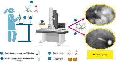
Abstract
Bacteriophages are viruses that infect bacteria. Researchers use different methods to study the characteristics of bacteriophages. Transmission electron microscope (TEM) is considered the best method to analyze these characteristics. However, the quality of TEM micrographs is significantly influenced by the preparation methods used to prepare the bacteriophages sample. In this study, researchers compared two different methods for preparing the bacteriophage samples. In one method was used SM buffer, while in the other used deionized water. The results were analyzed by TEM and compared with each other. Additionally, the viability of bacteriophage in deionized water and SM buffer at 4°C was determined through plaque assay within 72 hours. TEM micrographs showed that the quality of bacteriophage sample prepared with deionized water is superior to those prepared with SM buffer. Furthermore, the titer of the bacteriophages did not show a significant reduction during 72 hours in both SM and deionized water. In conclusion, the results suggested that preparation method can significantly impact the quality of TEM micrographs. Using sterile deionized water for the preparation of bacteriophages is a simple way to improve the quality of TEM micrographs and it is advisable to send the samples to the laboratory within 72 hours.
噬菌体是感染细菌的病毒。研究人员使用不同的方法来研究噬菌体的特征。透射电子显微镜(TEM)被认为是分析这些特征的最佳方法。然而,噬菌体样本的制备方法对 TEM 显微照片的质量有很大影响。在这项研究中,研究人员比较了两种不同的噬菌体样本制备方法。一种方法使用 SM 缓冲液,另一种方法使用去离子水。研究结果通过 TEM 进行分析并相互比较。此外,还通过斑块检测法测定了去离子水和 SM 缓冲液中的噬菌体在 4°C 温度下 72 小时内的存活率。TEM 显微照片显示,用去离子水制备的噬菌体样品质量优于用 SM 缓冲液制备的样品。此外,在 SM 和去离子水中 72 小时内,噬菌体的滴度都没有明显下降。总之,研究结果表明,制备方法会对 TEM 显微图像的质量产生重大影响。使用无菌去离子水制备噬菌体是提高 TEM 显微图片质量的一个简单方法,建议在 72 小时内将样品送至实验室。
期刊介绍:
The Journal of Virological Methods focuses on original, high quality research papers that describe novel and comprehensively tested methods which enhance human, animal, plant, bacterial or environmental virology and prions research and discovery.
The methods may include, but not limited to, the study of:
Viral components and morphology-
Virus isolation, propagation and development of viral vectors-
Viral pathogenesis, oncogenesis, vaccines and antivirals-
Virus replication, host-pathogen interactions and responses-
Virus transmission, prevention, control and treatment-
Viral metagenomics and virome-
Virus ecology, adaption and evolution-
Applied virology such as nanotechnology-
Viral diagnosis with novelty and comprehensive evaluation.
We seek articles, systematic reviews, meta-analyses and laboratory protocols that include comprehensive technical details with statistical confirmations that provide validations against current best practice, international standards or quality assurance programs and which advance knowledge in virology leading to improved medical, veterinary or agricultural practices and management.


 分享
分享
 求助内容:
求助内容: 应助结果提醒方式:
 扫码关注我们
