Junqing Yang, Pei Xu, Siyi Wu, Zhou Chen, Shiyan Fang, Haibo Xiao, Fengqing Hu, Lianyong Jiang, Lei Wang, Bin Mo, Fangbao Ding, Linley Li Lin, Jian Ye
{"title":"Raman spectroscopy for esophageal tumor diagnosis and delineation using machine learning and the portable Raman spectrometer.","authors":"Junqing Yang, Pei Xu, Siyi Wu, Zhou Chen, Shiyan Fang, Haibo Xiao, Fengqing Hu, Lianyong Jiang, Lei Wang, Bin Mo, Fangbao Ding, Linley Li Lin, Jian Ye","doi":"10.1016/j.saa.2024.124461","DOIUrl":null,"url":null,"abstract":"<p><p>Esophageal cancer is one of the leading causes of cancer-related deaths worldwide. The identification of residual tumor tissues in the surgical margin of esophageal cancer is essential for the treatment and prognosis of cancer patients. But the current diagnostic methods, either pathological frozen section or paraffin section examination, are laborious, time-consuming, and inconvenient. Raman spectroscopy is a label-free and non-invasive analytical technique that provides molecular information with high specificity. Here, we report the use of a portable Raman system and machine learning algorithms to achieve accurate diagnosis of esophageal tumor tissue in surgically resected specimens. We tested five machine learning-based classification methods, including k-Nearest Neighbors, Adaptive Boosting, Random Forest, Principal Component Analysis-Linear Discriminant Analysis, and Support Vector Machine (SVM). Among them, SVM shows the highest accuracy (88.61 %) in classifying the esophageal tumor and normal tissues. The portable Raman system demonstrates robust measurements with an acceptable focal plane shift of up to 3 mm, which enables large-area Raman mapping on resected tissues. Based on this, we finally achieve successful Raman visualization of tumor boundaries on surgical margin specimens, and the Raman measurement time is less than 5 min. This work provides a robust, convenient, accurate, and cost-effective tool for the diagnosis of esophageal cancer tumors, advancing toward Raman-based clinical intraoperative applications.</p>","PeriodicalId":94213,"journal":{"name":"Spectrochimica acta. Part A, Molecular and biomolecular spectroscopy","volume":"317 ","pages":"124461"},"PeriodicalIF":4.6000,"publicationDate":"2024-09-05","publicationTypes":"Journal Article","fieldsOfStudy":null,"isOpenAccess":false,"openAccessPdf":"","citationCount":"0","resultStr":null,"platform":"Semanticscholar","paperid":null,"PeriodicalName":"Spectrochimica acta. Part A, Molecular and biomolecular spectroscopy","FirstCategoryId":"1085","ListUrlMain":"https://doi.org/10.1016/j.saa.2024.124461","RegionNum":0,"RegionCategory":null,"ArticlePicture":[],"TitleCN":null,"AbstractTextCN":null,"PMCID":null,"EPubDate":"2024/5/12 0:00:00","PubModel":"Epub","JCR":"","JCRName":"","Score":null,"Total":0}
引用次数: 0
Abstract
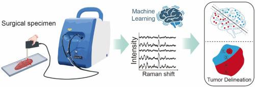
Esophageal cancer is one of the leading causes of cancer-related deaths worldwide. The identification of residual tumor tissues in the surgical margin of esophageal cancer is essential for the treatment and prognosis of cancer patients. But the current diagnostic methods, either pathological frozen section or paraffin section examination, are laborious, time-consuming, and inconvenient. Raman spectroscopy is a label-free and non-invasive analytical technique that provides molecular information with high specificity. Here, we report the use of a portable Raman system and machine learning algorithms to achieve accurate diagnosis of esophageal tumor tissue in surgically resected specimens. We tested five machine learning-based classification methods, including k-Nearest Neighbors, Adaptive Boosting, Random Forest, Principal Component Analysis-Linear Discriminant Analysis, and Support Vector Machine (SVM). Among them, SVM shows the highest accuracy (88.61 %) in classifying the esophageal tumor and normal tissues. The portable Raman system demonstrates robust measurements with an acceptable focal plane shift of up to 3 mm, which enables large-area Raman mapping on resected tissues. Based on this, we finally achieve successful Raman visualization of tumor boundaries on surgical margin specimens, and the Raman measurement time is less than 5 min. This work provides a robust, convenient, accurate, and cost-effective tool for the diagnosis of esophageal cancer tumors, advancing toward Raman-based clinical intraoperative applications.


 分享
分享

 求助内容:
求助内容: 应助结果提醒方式:
 扫码关注我们
