Serdar Kaymaz, Nilüfer Savurmuş, Uğur Karasu, Hüseyin Kaya, Furkan Ufuk, Ayşe Rüksan Ütebey, Veli Çobankara, Murat Yiğit
{"title":"Association between choroidal thickness and interstitial lung disease in patients with rheumatoid arthritis: A cross-sectional study.","authors":"Serdar Kaymaz, Nilüfer Savurmuş, Uğur Karasu, Hüseyin Kaya, Furkan Ufuk, Ayşe Rüksan Ütebey, Veli Çobankara, Murat Yiğit","doi":"10.46497/ArchRheumatol.2023.10116","DOIUrl":null,"url":null,"abstract":"<p><strong>Objectives: </strong>This study aimed to evaluate choroidal thickness (CT) in patients with rheumatoid arthritis (RA) and healthy controls and to determine its relationship with RA-associated interstitial lung disease (RA-ILD).</p><p><strong>Patients and methods: </strong>A total of 63 patients with RA and 36 age- and sex-matched healthy controls were recruited in the cross-sectional study. Serological findings, Disease Activity Score-28, disease duration, and medical treatment of patients were recorded. Patients with RA were subdivided into two groups: patients with RA-ILD (Group 1) and patients with RA but without ILD (RA-noILD; Group 2). CTs were measured using enhanced depth imaging optical coherence tomography. CT was measured at five points: the subfoveal region, 750 μm nasal and temporal to the fovea, 1500 μm nasal and temporal to the fovea. Patients with RA-ILD were evaluated with delta high-resolution computed tomography (ΔHRCT) and pulmonary function test to determine the severity of interstitial lung disease.</p><p><strong>Results: </strong>Four of 63 RA patients were excluded due to comorbidities. Thus, 59 RA patients, 20 in the RA-ILD group and 39 in the RA-noILD group, were included in the analyses. The RA groups were similar in terms of clinical characteristics and laboratory findings. There were statistically significant differences between Group 1, Group 2 and healthy controls (Group 3) compared to all CT values (p<0.05). The mean CT measured at 750 μm and 1500 μm nasal to the fovea was lowest in the RA-ILD group, followed by the RA-noILD and healthy groups (p<0.05). CT measurements did not correlate with the pulmonary function test and ΔHRCT.</p><p><strong>Conclusion: </strong>RA-ILD patients had a thinner CT measured at nasal points. However, there was no association between CT measurements and the severity of ILD.</p>","PeriodicalId":93884,"journal":{"name":"Archives of rheumatology","volume":"39 1","pages":"89-98"},"PeriodicalIF":1.1000,"publicationDate":"2023-08-18","publicationTypes":"Journal Article","fieldsOfStudy":null,"isOpenAccess":false,"openAccessPdf":"https://www.ncbi.nlm.nih.gov/pmc/articles/PMC11104761/pdf/","citationCount":"0","resultStr":null,"platform":"Semanticscholar","paperid":null,"PeriodicalName":"Archives of rheumatology","FirstCategoryId":"1085","ListUrlMain":"https://doi.org/10.46497/ArchRheumatol.2023.10116","RegionNum":0,"RegionCategory":null,"ArticlePicture":[],"TitleCN":null,"AbstractTextCN":null,"PMCID":null,"EPubDate":"2024/3/1 0:00:00","PubModel":"eCollection","JCR":"Q4","JCRName":"RHEUMATOLOGY","Score":null,"Total":0}
引用次数: 0
Abstract
Objectives: This study aimed to evaluate choroidal thickness (CT) in patients with rheumatoid arthritis (RA) and healthy controls and to determine its relationship with RA-associated interstitial lung disease (RA-ILD).
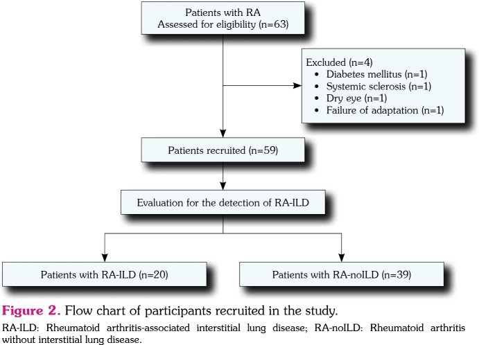
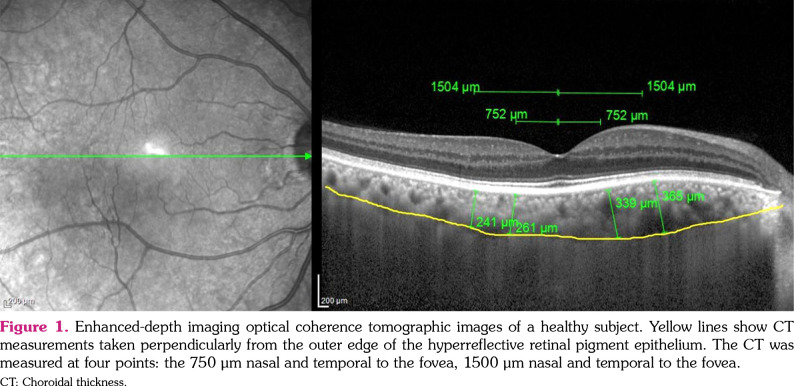
Patients and methods: A total of 63 patients with RA and 36 age- and sex-matched healthy controls were recruited in the cross-sectional study. Serological findings, Disease Activity Score-28, disease duration, and medical treatment of patients were recorded. Patients with RA were subdivided into two groups: patients with RA-ILD (Group 1) and patients with RA but without ILD (RA-noILD; Group 2). CTs were measured using enhanced depth imaging optical coherence tomography. CT was measured at five points: the subfoveal region, 750 μm nasal and temporal to the fovea, 1500 μm nasal and temporal to the fovea. Patients with RA-ILD were evaluated with delta high-resolution computed tomography (ΔHRCT) and pulmonary function test to determine the severity of interstitial lung disease.
Results: Four of 63 RA patients were excluded due to comorbidities. Thus, 59 RA patients, 20 in the RA-ILD group and 39 in the RA-noILD group, were included in the analyses. The RA groups were similar in terms of clinical characteristics and laboratory findings. There were statistically significant differences between Group 1, Group 2 and healthy controls (Group 3) compared to all CT values (p<0.05). The mean CT measured at 750 μm and 1500 μm nasal to the fovea was lowest in the RA-ILD group, followed by the RA-noILD and healthy groups (p<0.05). CT measurements did not correlate with the pulmonary function test and ΔHRCT.
Conclusion: RA-ILD patients had a thinner CT measured at nasal points. However, there was no association between CT measurements and the severity of ILD.


 分享
分享

 求助内容:
求助内容: 应助结果提醒方式:
 扫码关注我们
