Ductal carcinoma in situ develops within clonal fields of mutant cells in morphologically normal ducts
Stefan J Hutten, Hendrik A Messal, Esther H Lips, Michael Sheinman, Marta Ciwinska, Esmee Braams, Carolien van der Borden, Petra Kristel, Saskia Stoffers, Lodewyk FA Wessels, Grand Challenge PRECISION Consortium, Jos Jonkers, Jacco van Rheenen, Jelle Wesseling, Colinda LGJ Scheele
下载PDF
{"title":"Ductal carcinoma in situ develops within clonal fields of mutant cells in morphologically normal ducts","authors":"Stefan J Hutten, Hendrik A Messal, Esther H Lips, Michael Sheinman, Marta Ciwinska, Esmee Braams, Carolien van der Borden, Petra Kristel, Saskia Stoffers, Lodewyk FA Wessels, Grand Challenge PRECISION Consortium, Jos Jonkers, Jacco van Rheenen, Jelle Wesseling, Colinda LGJ Scheele","doi":"10.1002/path.6289","DOIUrl":null,"url":null,"abstract":"<p>Mutations are abundantly present in tissues of healthy individuals, including the breast epithelium. Yet it remains unknown whether mutant cells directly induce lesion formation or first spread, leading to a field of mutant cells that is predisposed towards lesion formation. To study the clonal and spatial relationships between morphologically normal breast epithelium adjacent to pre-cancerous lesions, we developed a three-dimensional (3D) imaging pipeline combined with spatially resolved genomics on archival, formalin-fixed breast tissue with the non-obligate breast cancer precursor ductal carcinoma <i>in situ</i> (DCIS). Using this 3D image-guided characterization method, we built high-resolution spatial maps of DNA copy number aberration (CNA) profiles within the DCIS lesion and the surrounding normal mammary ducts. We show that the local heterogeneity within a DCIS lesion is limited. However, by mapping the CNA profiles back onto the 3D reconstructed ductal subtree, we find that in eight out of 16 cases the healthy epithelium adjacent to the DCIS lesions has overlapping structural variations with the CNA profile of the DCIS. Together, our study indicates that pre-malignant breast transformations frequently develop within mutant clonal fields of morphologically normal-looking ducts. © 2024 The Authors. <i>The Journal of Pathology</i> published by John Wiley & Sons Ltd on behalf of The Pathological Society of Great Britain and Ireland.</p>","PeriodicalId":232,"journal":{"name":"The Journal of Pathology","volume":"263 3","pages":"360-371"},"PeriodicalIF":5.2000,"publicationDate":"2024-05-23","publicationTypes":"Journal Article","fieldsOfStudy":null,"isOpenAccess":false,"openAccessPdf":"https://onlinelibrary.wiley.com/doi/epdf/10.1002/path.6289","citationCount":"0","resultStr":null,"platform":"Semanticscholar","paperid":null,"PeriodicalName":"The Journal of Pathology","FirstCategoryId":"3","ListUrlMain":"https://pathsocjournals.onlinelibrary.wiley.com/doi/10.1002/path.6289","RegionNum":2,"RegionCategory":"医学","ArticlePicture":[],"TitleCN":null,"AbstractTextCN":null,"PMCID":null,"EPubDate":"","PubModel":"","JCR":"Q1","JCRName":"ONCOLOGY","Score":null,"Total":0}
引用次数: 0
引用
批量引用
Abstract
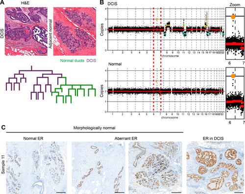
Mutations are abundantly present in tissues of healthy individuals, including the breast epithelium. Yet it remains unknown whether mutant cells directly induce lesion formation or first spread, leading to a field of mutant cells that is predisposed towards lesion formation. To study the clonal and spatial relationships between morphologically normal breast epithelium adjacent to pre-cancerous lesions, we developed a three-dimensional (3D) imaging pipeline combined with spatially resolved genomics on archival, formalin-fixed breast tissue with the non-obligate breast cancer precursor ductal carcinoma in situ (DCIS). Using this 3D image-guided characterization method, we built high-resolution spatial maps of DNA copy number aberration (CNA) profiles within the DCIS lesion and the surrounding normal mammary ducts. We show that the local heterogeneity within a DCIS lesion is limited. However, by mapping the CNA profiles back onto the 3D reconstructed ductal subtree, we find that in eight out of 16 cases the healthy epithelium adjacent to the DCIS lesions has overlapping structural variations with the CNA profile of the DCIS. Together, our study indicates that pre-malignant breast transformations frequently develop within mutant clonal fields of morphologically normal-looking ducts. © 2024 The Authors. The Journal of Pathology published by John Wiley & Sons Ltd on behalf of The Pathological Society of Great Britain and Ireland.
乳腺导管原位癌发生在形态正常的导管中突变细胞的克隆区内。
突变细胞大量存在于健康人的组织中,包括乳腺上皮细胞。然而,突变细胞是直接诱导病变形成,还是首先扩散,导致突变细胞场易形成病变,目前仍不得而知。为了研究与癌前病变相邻的形态正常的乳腺上皮细胞之间的克隆和空间关系,我们开发了一种三维(3D)成像流水线,结合空间分辨基因组学技术,用于研究福尔马林固定的存档乳腺组织和非可忽略的乳腺癌前体导管原位癌(DCIS)。利用这种三维图像引导的表征方法,我们建立了 DCIS 病变和周围正常乳腺导管内 DNA 拷贝数畸变 (CNA) 的高分辨率空间图谱。我们发现,DCIS 病灶内的局部异质性是有限的。然而,通过将 CNA 图谱映射回三维重建的乳腺导管子树,我们发现在 16 个病例中,有 8 个病例中邻近 DCIS 病变的健康上皮与 DCIS 的 CNA 图谱有重叠的结构变异。总之,我们的研究表明,乳腺恶变前病变经常发生在形态正常的导管突变克隆区内。© 2024 作者。病理学杂志》由约翰威利父子有限公司代表大不列颠及爱尔兰病理学会出版。
本文章由计算机程序翻译,如有差异,请以英文原文为准。


