Ariel Kerpel, Elizabeth Davenport, Amy L Proskovec, Yin Xi, Jarett D Berry, Zerrin Yetkin, Joseph Maldjian, Fang F Yu
{"title":"Antidepressant-related microstructural changes in the external capsule.","authors":"Ariel Kerpel, Elizabeth Davenport, Amy L Proskovec, Yin Xi, Jarett D Berry, Zerrin Yetkin, Joseph Maldjian, Fang F Yu","doi":"10.1007/s11682-024-00891-w","DOIUrl":null,"url":null,"abstract":"<p><p>Several magnetic resonance imaging (MRI) studies have reported that antidepressant medications are strongly linked to brain microstructural alterations. Notably, external capsule alterations have been reported to be a biological marker for therapeutic response. However, prior studies did not investigate whether a change in the neurite density or directional coherence of white matter (WM) fibers underlies the observed microstructural alterations. This MRI-based case-control study examined the relationship between patients' current use of antidepressant medications and advanced measurements of external capsule WM microstructure derived from multishell diffusion imaging using neurite orientation dispersion and density imaging (NODDI). The study compared a group of thirty-five participants who were taking antidepressant medications comprising selective serotonin reuptake inhibitors (SSRIs) (n = 25) and serotonin and norepinephrine reuptake inhibitors (SNRIs) with a control group of thirty-five individuals matched in terms of age, sex, race, and atherosclerotic cardiovascular risk factors. All participants were selected from the Dallas Heart Study phase 2, a multi-ethnic, population-based cohort study. A series of multiple linear regression analyses were conducted to predict microstructural characteristics of the bilateral external capsule using age, sex, and antidepressant medications as predictor variables. There was significantly reduced neurite density in the bilateral external capsules of patients taking SSRIs. Increased orientation dispersion in the external capsule was predominantly seen in patients taking SNRIs. Our findings suggest an association between specific external capsule microstructural changes and antidepressant medications, including reduced neurite density for SSRIs and increased orientation dispersion for SNRIs.</p>","PeriodicalId":9192,"journal":{"name":"Brain Imaging and Behavior","volume":" ","pages":"1044-1051"},"PeriodicalIF":2.4000,"publicationDate":"2024-10-01","publicationTypes":"Journal Article","fieldsOfStudy":null,"isOpenAccess":false,"openAccessPdf":"","citationCount":"0","resultStr":null,"platform":"Semanticscholar","paperid":null,"PeriodicalName":"Brain Imaging and Behavior","FirstCategoryId":"3","ListUrlMain":"https://doi.org/10.1007/s11682-024-00891-w","RegionNum":3,"RegionCategory":"医学","ArticlePicture":[],"TitleCN":null,"AbstractTextCN":null,"PMCID":null,"EPubDate":"2024/5/30 0:00:00","PubModel":"Epub","JCR":"Q2","JCRName":"NEUROIMAGING","Score":null,"Total":0}
引用次数: 0
Abstract
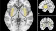
Several magnetic resonance imaging (MRI) studies have reported that antidepressant medications are strongly linked to brain microstructural alterations. Notably, external capsule alterations have been reported to be a biological marker for therapeutic response. However, prior studies did not investigate whether a change in the neurite density or directional coherence of white matter (WM) fibers underlies the observed microstructural alterations. This MRI-based case-control study examined the relationship between patients' current use of antidepressant medications and advanced measurements of external capsule WM microstructure derived from multishell diffusion imaging using neurite orientation dispersion and density imaging (NODDI). The study compared a group of thirty-five participants who were taking antidepressant medications comprising selective serotonin reuptake inhibitors (SSRIs) (n = 25) and serotonin and norepinephrine reuptake inhibitors (SNRIs) with a control group of thirty-five individuals matched in terms of age, sex, race, and atherosclerotic cardiovascular risk factors. All participants were selected from the Dallas Heart Study phase 2, a multi-ethnic, population-based cohort study. A series of multiple linear regression analyses were conducted to predict microstructural characteristics of the bilateral external capsule using age, sex, and antidepressant medications as predictor variables. There was significantly reduced neurite density in the bilateral external capsules of patients taking SSRIs. Increased orientation dispersion in the external capsule was predominantly seen in patients taking SNRIs. Our findings suggest an association between specific external capsule microstructural changes and antidepressant medications, including reduced neurite density for SSRIs and increased orientation dispersion for SNRIs.
期刊介绍:
Brain Imaging and Behavior is a bi-monthly, peer-reviewed journal, that publishes clinically relevant research using neuroimaging approaches to enhance our understanding of disorders of higher brain function. The journal is targeted at clinicians and researchers in fields concerned with human brain-behavior relationships, such as neuropsychology, psychiatry, neurology, neurosurgery, rehabilitation, and cognitive neuroscience.


 分享
分享
 求助内容:
求助内容: 应助结果提醒方式:
 扫码关注我们
