Diana Romenskaja, Ugnė Jonavičė, Augustas Pivoriūnas
{"title":"Extracellular vesicles promote autophagy in human microglia through lipid raft-dependent mechanisms","authors":"Diana Romenskaja, Ugnė Jonavičė, Augustas Pivoriūnas","doi":"10.1111/febs.17192","DOIUrl":null,"url":null,"abstract":"<p>Autophagy dysfunction has been closely related with pathogenesis of many neurodegenerative diseases and therefore represents a potential therapeutic target. Extracellular vesicles (EVs) may act as potent anti-inflammatory agents and also modulators of autophagy in target cells. However, the molecular mechanisms by which EVs modulate autophagy flux in human microglia remain largely unexplored. In the present study, we investigated the effects of EVs derived from human oral mucosa stem cells on the autophagy in human microglia. We demonstrate that EVs promoted autophagy and autophagic flux in human microglia and that this process was dependent on the integrity of lipid rafts. Lipopolysaccharide (LPS) also activated autophagy, but combined treatment with EVs and LPS suppressed autophagy response, indicating interference between these signaling pathways. Blockage of Toll-like receptor 4 (TLR4) with anti-TLR4 antibody suppressed EV-induced autophagy. Furthermore, inhibition of the EV-associated heat shock protein (HSP70) chaperone which is one of the endogenous ligands of the TLR4 also suppressed EV-induced lipid raft formation and autophagy. Pre-treatment of microglia with a selective inhibitor of αvβ3/αvβ5 integrins cilengitide inhibited EV-induced autophagy. Finally, blockage of purinergic P2X4 receptor (P2X4R) with selective inhibitor 5-BDBD also suppressed EV-induced autophagy. In conclusion, we demonstrate that EVs activate autophagy in human microglia through interaction with HSP70/TLR4, αVβ3/αVβ5, and P2X4R signaling pathways and that these effects depend on the integrity of lipid rafts. Our findings could be used to develop new therapeutic strategies targeting disease-associated microglia.</p>","PeriodicalId":94226,"journal":{"name":"The FEBS journal","volume":"291 16","pages":"3706-3722"},"PeriodicalIF":4.2000,"publicationDate":"2024-06-05","publicationTypes":"Journal Article","fieldsOfStudy":null,"isOpenAccess":false,"openAccessPdf":"https://onlinelibrary.wiley.com/doi/epdf/10.1111/febs.17192","citationCount":"0","resultStr":null,"platform":"Semanticscholar","paperid":null,"PeriodicalName":"The FEBS journal","FirstCategoryId":"1085","ListUrlMain":"https://febs.onlinelibrary.wiley.com/doi/10.1111/febs.17192","RegionNum":0,"RegionCategory":null,"ArticlePicture":[],"TitleCN":null,"AbstractTextCN":null,"PMCID":null,"EPubDate":"","PubModel":"","JCR":"","JCRName":"","Score":null,"Total":0}
引用次数: 0
Abstract
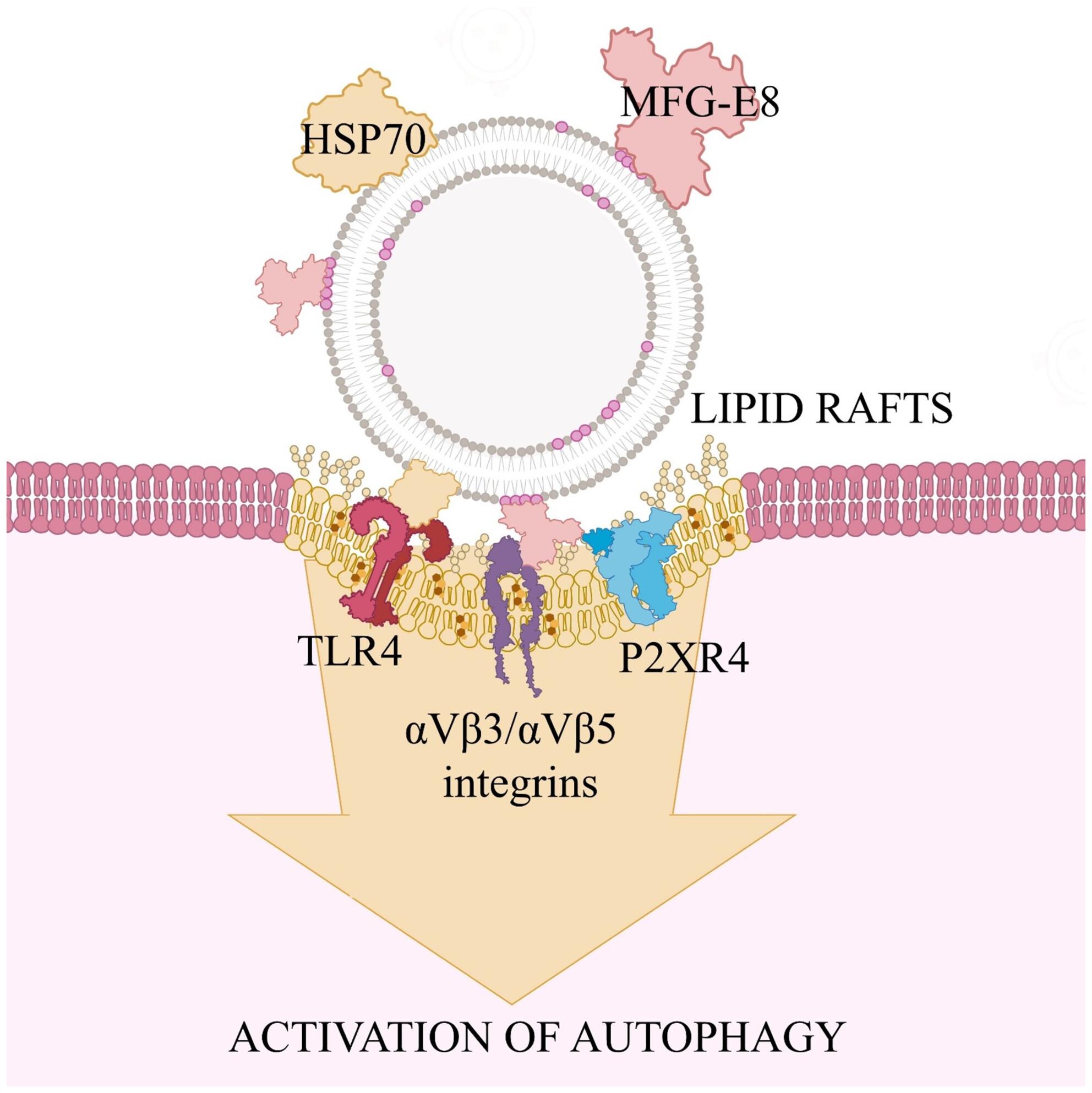
Autophagy dysfunction has been closely related with pathogenesis of many neurodegenerative diseases and therefore represents a potential therapeutic target. Extracellular vesicles (EVs) may act as potent anti-inflammatory agents and also modulators of autophagy in target cells. However, the molecular mechanisms by which EVs modulate autophagy flux in human microglia remain largely unexplored. In the present study, we investigated the effects of EVs derived from human oral mucosa stem cells on the autophagy in human microglia. We demonstrate that EVs promoted autophagy and autophagic flux in human microglia and that this process was dependent on the integrity of lipid rafts. Lipopolysaccharide (LPS) also activated autophagy, but combined treatment with EVs and LPS suppressed autophagy response, indicating interference between these signaling pathways. Blockage of Toll-like receptor 4 (TLR4) with anti-TLR4 antibody suppressed EV-induced autophagy. Furthermore, inhibition of the EV-associated heat shock protein (HSP70) chaperone which is one of the endogenous ligands of the TLR4 also suppressed EV-induced lipid raft formation and autophagy. Pre-treatment of microglia with a selective inhibitor of αvβ3/αvβ5 integrins cilengitide inhibited EV-induced autophagy. Finally, blockage of purinergic P2X4 receptor (P2X4R) with selective inhibitor 5-BDBD also suppressed EV-induced autophagy. In conclusion, we demonstrate that EVs activate autophagy in human microglia through interaction with HSP70/TLR4, αVβ3/αVβ5, and P2X4R signaling pathways and that these effects depend on the integrity of lipid rafts. Our findings could be used to develop new therapeutic strategies targeting disease-associated microglia.


 分享
分享

 求助内容:
求助内容: 应助结果提醒方式:
 扫码关注我们
