Amygdala enlargement can occur in temporal lobe epilepsy, and increased amygdala volume is also reported in sudden unexpected death in epilepsy (SUDEP). Apnea can be induced by amygdala stimulation, and postconvulsive central apnea (PCCA) and generalized seizures are both known SUDEP risk factors. Neurite orientation dispersion and density imaging (NODDI) has recently provided additional information on altered amygdala microstructure in SUDEP. In a series of 24 surgical temporal lobe epilepsy cases, our aim was to quantify amygdala cellular pathology parameters that could predict enlargement, NODDI changes, and ictal respiratory dysfunction.
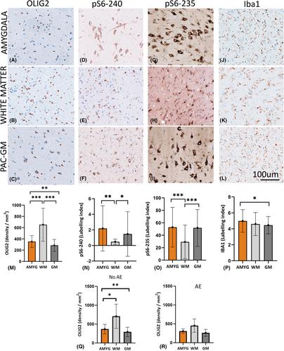
Using whole slide scanning automated quantitative image analysis methods, parallel evaluation of myelin, axons, dendrites, oligodendroglia, microglia, astroglia, neurons, serotonergic networks, mTOR-pathway activation (pS6) and phosphorylated tau (pTau; AT8, AT100, PHF) in amygdala, periamygdala cortex, and white matter regions of interest were compared with preoperative magnetic resonance imaging data on amygdala size, and in 13 cases with NODDI and evidence of ictal-associated apnea.
We observed significantly higher glial labeling (Iba1, glial fibrillary acidic protein, Olig2) in amygdala regions compared to cortex and a strong positive correlation between Olig2 and Iba1 in the amygdala. Larger amygdala volumes correlated with lower microtubule-associated protein (MAP2), whereas higher NODDI orientation dispersion index correlated with lower Olig2 cell densities. In the three cases with recorded PCCA, higher MAP2 and pS6-235 expression was noted than in those without. pTau did not correlate with SUDEP risk factors, including seizure frequency.
Histological quantitation of amygdala microstructure can shed light on enlargement and diffusion imaging alterations in epilepsy to explore possible mechanisms of amygdala dysfunction, including mTOR pathway activation, that in turn may increase the risk for SUDEP.


 分享
分享
 求助内容:
求助内容: 应助结果提醒方式:
 扫码关注我们
