Meltem Ivren, Asan Cherkezov, David Reuss, Daniel Haux, Christel Herold-Mende, Alexander Mohr, Sandro M Krieg, Andreas Unterberg, Alexander Younsi
{"title":"Intracranial angioleiomyoma: a case series of seven patients and review of the literature.","authors":"Meltem Ivren, Asan Cherkezov, David Reuss, Daniel Haux, Christel Herold-Mende, Alexander Mohr, Sandro M Krieg, Andreas Unterberg, Alexander Younsi","doi":"10.1007/s11060-024-04734-y","DOIUrl":null,"url":null,"abstract":"<p><strong>Purpose: </strong>Angioleiomyoma, predominantly arising from the extremities, is a benign soft tissue tumor. Reports on its intracranial location are rare. We assessed clinical, radiological, and pathological features of intracranial angioleiomyoma (iALM) treated at our neurosurgical institution.</p><p><strong>Methods: </strong>We consecutively enrolled all patients with neuropathologically confirmed iALM treated at a single neurosurgical institution between 2013 and 2021. Clinical and imaging data were collected, and histological tissue sections were analyzed. A review of the literature on iALM was conducted.</p><p><strong>Results: </strong>Seven patients with iALM (four female) with a median age of 45 years (range: 32-76 years) were identified. In three cases, the lesion was found incidentally. In magnetic resonance imaging (MRI), all tumors were hypo- to isointense on T1-weighted, hyperintense on T2-weighted sequences, and gadolinium-enhancing. A strong FLAIR signal was seen in six patients. Surgery consisted of gross total resection in all cases without perioperative complications. Neuropathological staining was positive for smooth muscle actin (SMA) in all lesions. Mature smooth muscle cells arranged around blood vessels were typically observed. The Ki-67 index was ≤ 3%. The patients were discharged after a median of 6 days (range: 4-9 days). During a median follow-up time of 14 months (range: 4-41 months), no tumor recurrence occurred. In the current literature, 42 additional cases of iALM were identified.</p><p><strong>Conclusion: </strong>Intracranial angioleiomyoma is a benign soft tissue tumor treated by gross total resection. Tumor morphology and positive staining for SMA lead to the neuropathological diagnosis.</p>","PeriodicalId":16425,"journal":{"name":"Journal of Neuro-Oncology","volume":null,"pages":null},"PeriodicalIF":3.2000,"publicationDate":"2024-09-01","publicationTypes":"Journal Article","fieldsOfStudy":null,"isOpenAccess":false,"openAccessPdf":"https://www.ncbi.nlm.nih.gov/pmc/articles/PMC11341739/pdf/","citationCount":"0","resultStr":null,"platform":"Semanticscholar","paperid":null,"PeriodicalName":"Journal of Neuro-Oncology","FirstCategoryId":"3","ListUrlMain":"https://doi.org/10.1007/s11060-024-04734-y","RegionNum":2,"RegionCategory":"医学","ArticlePicture":[],"TitleCN":null,"AbstractTextCN":null,"PMCID":null,"EPubDate":"2024/6/6 0:00:00","PubModel":"Epub","JCR":"Q2","JCRName":"CLINICAL NEUROLOGY","Score":null,"Total":0}
引用次数: 0
Abstract
Purpose: Angioleiomyoma, predominantly arising from the extremities, is a benign soft tissue tumor. Reports on its intracranial location are rare. We assessed clinical, radiological, and pathological features of intracranial angioleiomyoma (iALM) treated at our neurosurgical institution.
Methods: We consecutively enrolled all patients with neuropathologically confirmed iALM treated at a single neurosurgical institution between 2013 and 2021. Clinical and imaging data were collected, and histological tissue sections were analyzed. A review of the literature on iALM was conducted.
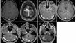
Results: Seven patients with iALM (four female) with a median age of 45 years (range: 32-76 years) were identified. In three cases, the lesion was found incidentally. In magnetic resonance imaging (MRI), all tumors were hypo- to isointense on T1-weighted, hyperintense on T2-weighted sequences, and gadolinium-enhancing. A strong FLAIR signal was seen in six patients. Surgery consisted of gross total resection in all cases without perioperative complications. Neuropathological staining was positive for smooth muscle actin (SMA) in all lesions. Mature smooth muscle cells arranged around blood vessels were typically observed. The Ki-67 index was ≤ 3%. The patients were discharged after a median of 6 days (range: 4-9 days). During a median follow-up time of 14 months (range: 4-41 months), no tumor recurrence occurred. In the current literature, 42 additional cases of iALM were identified.
Conclusion: Intracranial angioleiomyoma is a benign soft tissue tumor treated by gross total resection. Tumor morphology and positive staining for SMA lead to the neuropathological diagnosis.
期刊介绍:
The Journal of Neuro-Oncology is a multi-disciplinary journal encompassing basic, applied, and clinical investigations in all research areas as they relate to cancer and the central nervous system. It provides a single forum for communication among neurologists, neurosurgeons, radiotherapists, medical oncologists, neuropathologists, neurodiagnosticians, and laboratory-based oncologists conducting relevant research. The Journal of Neuro-Oncology does not seek to isolate the field, but rather to focus the efforts of many disciplines in one publication through a format which pulls together these diverse interests. More than any other field of oncology, cancer of the central nervous system requires multi-disciplinary approaches. To alleviate having to scan dozens of journals of cell biology, pathology, laboratory and clinical endeavours, JNO is a periodical in which current, high-quality, relevant research in all aspects of neuro-oncology may be found.


 分享
分享
 求助内容:
求助内容: 应助结果提醒方式:
 扫码关注我们
