Lourenço Galizia Heitzmann, Igel de Souza Aquino, Antonio Carlos Tenor Junior, Miguel Pereira da Costa, Monica Paschoal Nogueira
{"title":"Anatomical study of the safety corridor for bridge plating positioned on the lateral border of the humerus.","authors":"Lourenço Galizia Heitzmann, Igel de Souza Aquino, Antonio Carlos Tenor Junior, Miguel Pereira da Costa, Monica Paschoal Nogueira","doi":"10.1007/s00276-024-03405-x","DOIUrl":null,"url":null,"abstract":"<p><strong>Purpose: </strong>This study shows the danger zone and the safety corridor in the lateral approach with bridge plating by measuring the distance between the lateral side of the plate positioned on the lateral aspect of the humerus and the radial nerve after it pierces the lateral intermuscular septum, in the different forearm positions.</p><p><strong>Methods: </strong>Forty arms of 20 human cadavers were used, the radial nerve was identified and marked on the lateral surface the radial nerve at the exit of the lateral intermuscular septum and anteriorisation of the nerve in relation to the humeral shaft and the lateral epicondyle was also marked. The distances were measured with a digital caliper. A submuscular extraperiosteal corridor was created, proximally between the biceps brachialis and deltoid muscle and distally between the triceps and brachioradialis muscle, followed by the positioning of the low contact large fragments contoured plate with 14 combined holes (fixed and cortical angle), inserted from distal to proximal. Measurements were performed in four positions (elbow flexion with forearm pronation, elbow flexion with forearm supination, elbow extension with forearm pronation and elbow extension with forearm supination).</p><p><strong>Results: </strong>Significant statistical differences occurred with the different positions, and the elbow flexion with forearm supination was shown to be the position that provides the safest submuscular extraperiosteal corridor in a lateral approach of the humerus.</p><p><strong>Conclusion: </strong>The danger zone of radial nerve is an area that extends from 15 cm to 5 cm proximal to the lateral epicondyle and the safest way to create a submuscular and extraperiosteal corridor in the lateral region of the humerus is with the elbow in flexion and the forearm in supination.</p>","PeriodicalId":49461,"journal":{"name":"Surgical and Radiologic Anatomy","volume":" ","pages":"1439-1445"},"PeriodicalIF":1.2000,"publicationDate":"2024-09-01","publicationTypes":"Journal Article","fieldsOfStudy":null,"isOpenAccess":false,"openAccessPdf":"","citationCount":"0","resultStr":null,"platform":"Semanticscholar","paperid":null,"PeriodicalName":"Surgical and Radiologic Anatomy","FirstCategoryId":"3","ListUrlMain":"https://doi.org/10.1007/s00276-024-03405-x","RegionNum":4,"RegionCategory":"医学","ArticlePicture":[],"TitleCN":null,"AbstractTextCN":null,"PMCID":null,"EPubDate":"2024/6/10 0:00:00","PubModel":"Epub","JCR":"Q2","JCRName":"Medicine","Score":null,"Total":0}
引用次数: 0
Abstract
Purpose: This study shows the danger zone and the safety corridor in the lateral approach with bridge plating by measuring the distance between the lateral side of the plate positioned on the lateral aspect of the humerus and the radial nerve after it pierces the lateral intermuscular septum, in the different forearm positions.
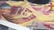
Methods: Forty arms of 20 human cadavers were used, the radial nerve was identified and marked on the lateral surface the radial nerve at the exit of the lateral intermuscular septum and anteriorisation of the nerve in relation to the humeral shaft and the lateral epicondyle was also marked. The distances were measured with a digital caliper. A submuscular extraperiosteal corridor was created, proximally between the biceps brachialis and deltoid muscle and distally between the triceps and brachioradialis muscle, followed by the positioning of the low contact large fragments contoured plate with 14 combined holes (fixed and cortical angle), inserted from distal to proximal. Measurements were performed in four positions (elbow flexion with forearm pronation, elbow flexion with forearm supination, elbow extension with forearm pronation and elbow extension with forearm supination).
Results: Significant statistical differences occurred with the different positions, and the elbow flexion with forearm supination was shown to be the position that provides the safest submuscular extraperiosteal corridor in a lateral approach of the humerus.
Conclusion: The danger zone of radial nerve is an area that extends from 15 cm to 5 cm proximal to the lateral epicondyle and the safest way to create a submuscular and extraperiosteal corridor in the lateral region of the humerus is with the elbow in flexion and the forearm in supination.
期刊介绍:
Anatomy is a morphological science which cannot fail to interest the clinician. The practical application of anatomical research to clinical problems necessitates special adaptation and selectivity in choosing from numerous international works. Although there is a tendency to believe that meaningful advances in anatomy are unlikely, constant revision is necessary. Surgical and Radiologic Anatomy, the first international journal of Clinical anatomy has been created in this spirit.
Its goal is to serve clinicians, regardless of speciality-physicians, surgeons, radiologists or other specialists-as an indispensable aid with which they can improve their knowledge of anatomy. Each issue includes: Original papers, review articles, articles on the anatomical bases of medical, surgical and radiological techniques, articles of normal radiologic anatomy, brief reviews of anatomical publications of clinical interest.
Particular attention is given to high quality illustrations, which are indispensable for a better understanding of anatomical problems.
Surgical and Radiologic Anatomy is a journal written by anatomists for clinicians with a special interest in anatomy.


 分享
分享
 求助内容:
求助内容: 应助结果提醒方式:
 扫码关注我们
