Mahsa Soti Khiabani, Maryam Monajemzadeh, Hojatollah Raji, Fatemeh Zamani, Mohammad Vaseie, Neda Pak
{"title":"Pediatric Inflammatory Myofibroblastic Tumor of Rectosigmoid Junction: A Case Report and Review of the Literature.","authors":"Mahsa Soti Khiabani, Maryam Monajemzadeh, Hojatollah Raji, Fatemeh Zamani, Mohammad Vaseie, Neda Pak","doi":"10.30699/ijp.2024.2003653.3122","DOIUrl":null,"url":null,"abstract":"<p><p>The occurrence of rectosigmoid junction inflammatory myofibroblastic tumor (IMT) is uncommon in children. This is a rare form of mesenchymal tumor, belonging to the category of soft tissue tumors, and can be found at any anatomical site from the central nervous system to the gastrointestinal tract. Our patient was a 10-year-old male subject complaining of lack of defecation and constipation. The patient had decreased the frequency of defecation and constipation about two weeks before his referral and had not improved despite the use of laxatives. The abdomen was completely distended and there was no tenderness or guarding in the examination. Several airfluid levels are shown on the abdominal X-ray. In the ultrasound, free fluid was reported in the interlobular and pelvic spaces. The patient was transferred into the operating room. A tumor of the rectosigmoid junction was detected. Histopathologic studies showed evidence of IMT. IMT is a rare neoplasm of unknown origin, which may occur in various sites of the body. Complete surgical removal is usually curative, but early detection of recurrence is required. Treatment options include chemotherapy, radiation therapy, and immunotherapy. Further investigations are needed to improve the understanding and management of this rare tumor.</p>","PeriodicalId":38900,"journal":{"name":"Iranian Journal of Pathology","volume":"19 1","pages":"132-136"},"PeriodicalIF":0.0000,"publicationDate":"2024-01-01","publicationTypes":"Journal Article","fieldsOfStudy":null,"isOpenAccess":false,"openAccessPdf":"https://www.ncbi.nlm.nih.gov/pmc/articles/PMC11164313/pdf/","citationCount":"0","resultStr":null,"platform":"Semanticscholar","paperid":null,"PeriodicalName":"Iranian Journal of Pathology","FirstCategoryId":"1085","ListUrlMain":"https://doi.org/10.30699/ijp.2024.2003653.3122","RegionNum":0,"RegionCategory":null,"ArticlePicture":[],"TitleCN":null,"AbstractTextCN":null,"PMCID":null,"EPubDate":"2023/12/29 0:00:00","PubModel":"Epub","JCR":"Q3","JCRName":"Medicine","Score":null,"Total":0}
引用次数: 0
Abstract
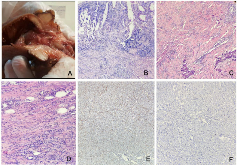
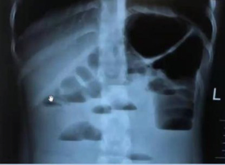
The occurrence of rectosigmoid junction inflammatory myofibroblastic tumor (IMT) is uncommon in children. This is a rare form of mesenchymal tumor, belonging to the category of soft tissue tumors, and can be found at any anatomical site from the central nervous system to the gastrointestinal tract. Our patient was a 10-year-old male subject complaining of lack of defecation and constipation. The patient had decreased the frequency of defecation and constipation about two weeks before his referral and had not improved despite the use of laxatives. The abdomen was completely distended and there was no tenderness or guarding in the examination. Several airfluid levels are shown on the abdominal X-ray. In the ultrasound, free fluid was reported in the interlobular and pelvic spaces. The patient was transferred into the operating room. A tumor of the rectosigmoid junction was detected. Histopathologic studies showed evidence of IMT. IMT is a rare neoplasm of unknown origin, which may occur in various sites of the body. Complete surgical removal is usually curative, but early detection of recurrence is required. Treatment options include chemotherapy, radiation therapy, and immunotherapy. Further investigations are needed to improve the understanding and management of this rare tumor.
直肠乙状结肠交界处炎性肌纤维母细胞瘤(IMT)在儿童中并不常见。这是一种罕见的间叶肿瘤,属于软组织肿瘤,可发生在从中枢神经系统到胃肠道的任何解剖部位。我们的患者是一名 10 岁男性,主诉排便不畅和便秘。在转诊前两周左右,患者的排便和便秘次数减少,尽管使用了泻药,但情况仍未改善。腹部完全胀满,检查时没有触痛或压迫感。腹部 X 光片显示有多处积气。超声波检查显示,小叶间隙和骨盆间隙有游离液体。患者被转入手术室。发现直肠乙状结肠交界处有肿瘤。组织病理学研究显示,该肿瘤为IMT。IMT是一种原因不明的罕见肿瘤,可能发生在身体的不同部位。完全手术切除通常可以治愈,但需要及早发现复发。治疗方法包括化疗、放疗和免疫疗法。为了更好地了解和治疗这种罕见肿瘤,还需要进一步的研究。


 分享
分享
 求助内容:
求助内容: 应助结果提醒方式:
 扫码关注我们
