Alfonso Estudillo Romero, Raffaella Migliaccio, Bénédicte Batrancourt, Pierre Jannin, John S H Baxter
{"title":"Analysis of convolutional neural networks for fronto-temporal dementia biomarker discovery.","authors":"Alfonso Estudillo Romero, Raffaella Migliaccio, Bénédicte Batrancourt, Pierre Jannin, John S H Baxter","doi":"10.1007/s11548-024-03197-w","DOIUrl":null,"url":null,"abstract":"<p><strong>Purpose: </strong>Frontotemporal lobe dementia (FTD) results from the degeneration of the frontal and temporal lobes. It can manifest in several different ways, leading to the definition of variants characterised by their distinctive symptomatologies. As these variants are detected based on their symptoms, it can be unclear if they represent different types of FTD or different symptomatological axes. The goal of this paper is to investigate this question with a constrained cohort of FTD patients in order to see if the heterogeneity within this cohort can be inferred from medical images rather than symptom severity measurements.</p><p><strong>Methods: </strong>An ensemble of convolutional neural networks (CNNs) is used to classify diffusion tensor images collected from two databases consisting of 72 patients with behavioural variant FTD and 120 healthy controls. FTD biomarkers were found using voxel-based analysis on the sensitivities of these CNNs. Sparse principal components analysis (sPCA) is then applied on the sensitivities arising from the patient cohort in order to identify the axes along which the patients express these biomarkers. Finally, this is correlated with their symptom severity measurements in order to interpret the clinical presentation of each axis.</p><p><strong>Results: </strong>The CNNs result in sensitivities and specificities between 83 and 92%. As expected, our analysis determines that all the robust biomarkers arise from the frontal and temporal lobes. sPCA identified four axes in terms of biomarker expression which are correlated with symptom severity measurements.</p><p><strong>Conclusion: </strong>Our analysis confirms that behavioural variant FTD is not a singular type or spectrum of FTD, but rather that it has multiple symptomatological axes that relate to distinct regions of the frontal and temporal lobes. This analysis suggests that medical images can be used to understand the heterogeneity of FTD patients and the underlying anatomical changes that lead to their different clinical presentations.</p>","PeriodicalId":51251,"journal":{"name":"International Journal of Computer Assisted Radiology and Surgery","volume":" ","pages":"2339-2349"},"PeriodicalIF":2.3000,"publicationDate":"2024-12-01","publicationTypes":"Journal Article","fieldsOfStudy":null,"isOpenAccess":false,"openAccessPdf":"","citationCount":"0","resultStr":null,"platform":"Semanticscholar","paperid":null,"PeriodicalName":"International Journal of Computer Assisted Radiology and Surgery","FirstCategoryId":"5","ListUrlMain":"https://doi.org/10.1007/s11548-024-03197-w","RegionNum":3,"RegionCategory":"医学","ArticlePicture":[],"TitleCN":null,"AbstractTextCN":null,"PMCID":null,"EPubDate":"2024/6/14 0:00:00","PubModel":"Epub","JCR":"Q3","JCRName":"ENGINEERING, BIOMEDICAL","Score":null,"Total":0}
引用次数: 0
Abstract
Purpose: Frontotemporal lobe dementia (FTD) results from the degeneration of the frontal and temporal lobes. It can manifest in several different ways, leading to the definition of variants characterised by their distinctive symptomatologies. As these variants are detected based on their symptoms, it can be unclear if they represent different types of FTD or different symptomatological axes. The goal of this paper is to investigate this question with a constrained cohort of FTD patients in order to see if the heterogeneity within this cohort can be inferred from medical images rather than symptom severity measurements.
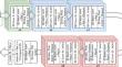
Methods: An ensemble of convolutional neural networks (CNNs) is used to classify diffusion tensor images collected from two databases consisting of 72 patients with behavioural variant FTD and 120 healthy controls. FTD biomarkers were found using voxel-based analysis on the sensitivities of these CNNs. Sparse principal components analysis (sPCA) is then applied on the sensitivities arising from the patient cohort in order to identify the axes along which the patients express these biomarkers. Finally, this is correlated with their symptom severity measurements in order to interpret the clinical presentation of each axis.
Results: The CNNs result in sensitivities and specificities between 83 and 92%. As expected, our analysis determines that all the robust biomarkers arise from the frontal and temporal lobes. sPCA identified four axes in terms of biomarker expression which are correlated with symptom severity measurements.
Conclusion: Our analysis confirms that behavioural variant FTD is not a singular type or spectrum of FTD, but rather that it has multiple symptomatological axes that relate to distinct regions of the frontal and temporal lobes. This analysis suggests that medical images can be used to understand the heterogeneity of FTD patients and the underlying anatomical changes that lead to their different clinical presentations.
期刊介绍:
The International Journal for Computer Assisted Radiology and Surgery (IJCARS) is a peer-reviewed journal that provides a platform for closing the gap between medical and technical disciplines, and encourages interdisciplinary research and development activities in an international environment.


 分享
分享
 求助内容:
求助内容: 应助结果提醒方式:
 扫码关注我们
