Zachary Hoglund, Nancy Ruiz-Uribe, Eric del Sastre, Benjamin Woost, Elizabeth Bader, Joshua Bailey, Bradley T. Hyman, Theodore Zwang, Rachel E. Bennett
{"title":"Brain vasculature accumulates tau and is spatially related to tau tangle pathology in Alzheimer’s disease","authors":"Zachary Hoglund, Nancy Ruiz-Uribe, Eric del Sastre, Benjamin Woost, Elizabeth Bader, Joshua Bailey, Bradley T. Hyman, Theodore Zwang, Rachel E. Bennett","doi":"10.1007/s00401-024-02751-9","DOIUrl":null,"url":null,"abstract":"<div><p>Insoluble pathogenic proteins accumulate along blood vessels in conditions of cerebral amyloid angiopathy (CAA), exerting a toxic effect on vascular cells and impacting cerebral homeostasis. In this work, we provide new evidence from three-dimensional human brain histology that tau protein, the main component of neurofibrillary tangles, can similarly accumulate along brain vascular segments. We quantitatively assessed n = 6 Alzheimer’s disease (AD), and n = 6 normal aging control brains and saw that tau-positive blood vessel segments were present in all AD cases. Tau-positive vessels are enriched for tau at levels higher than the surrounding tissue and appear to affect arterioles across cortical layers (I–V). Further, vessels isolated from these AD tissues were enriched for N-terminal tau and tau phosphorylated at T181 and T217. Importantly, tau-positive vessels are associated with local areas of increased tau neurofibrillary tangles. This suggests that accumulation of tau around blood vessels may reflect a local clearance failure. In sum, these data indicate that tau, like amyloid beta, accumulates along blood vessels and may exert a significant influence on vasculature in the setting of AD.</p></div>","PeriodicalId":7012,"journal":{"name":"Acta Neuropathologica","volume":"147 1","pages":""},"PeriodicalIF":9.3000,"publicationDate":"2024-06-17","publicationTypes":"Journal Article","fieldsOfStudy":null,"isOpenAccess":false,"openAccessPdf":"https://www.ncbi.nlm.nih.gov/pmc/articles/PMC11182845/pdf/","citationCount":"0","resultStr":null,"platform":"Semanticscholar","paperid":null,"PeriodicalName":"Acta Neuropathologica","FirstCategoryId":"3","ListUrlMain":"https://link.springer.com/article/10.1007/s00401-024-02751-9","RegionNum":1,"RegionCategory":"医学","ArticlePicture":[],"TitleCN":null,"AbstractTextCN":null,"PMCID":null,"EPubDate":"","PubModel":"","JCR":"Q1","JCRName":"CLINICAL NEUROLOGY","Score":null,"Total":0}
引用次数: 0
Abstract
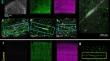
Insoluble pathogenic proteins accumulate along blood vessels in conditions of cerebral amyloid angiopathy (CAA), exerting a toxic effect on vascular cells and impacting cerebral homeostasis. In this work, we provide new evidence from three-dimensional human brain histology that tau protein, the main component of neurofibrillary tangles, can similarly accumulate along brain vascular segments. We quantitatively assessed n = 6 Alzheimer’s disease (AD), and n = 6 normal aging control brains and saw that tau-positive blood vessel segments were present in all AD cases. Tau-positive vessels are enriched for tau at levels higher than the surrounding tissue and appear to affect arterioles across cortical layers (I–V). Further, vessels isolated from these AD tissues were enriched for N-terminal tau and tau phosphorylated at T181 and T217. Importantly, tau-positive vessels are associated with local areas of increased tau neurofibrillary tangles. This suggests that accumulation of tau around blood vessels may reflect a local clearance failure. In sum, these data indicate that tau, like amyloid beta, accumulates along blood vessels and may exert a significant influence on vasculature in the setting of AD.
在脑淀粉样血管病(CAA)的情况下,不溶性致病蛋白会沿着血管积聚,对血管细胞产生毒性作用,影响大脑的稳态。在这项工作中,我们从三维人脑组织学中提供了新的证据,证明神经纤维缠结的主要成分 tau 蛋白同样会沿着脑血管节段积聚。我们对 n = 6 个阿尔茨海默病(AD)病例和 n = 6 个正常衰老对照组大脑进行了定量评估,发现所有 AD 病例中都存在 tau 蛋白阳性的血管片段。Tau阳性血管的tau含量高于周围组织,而且似乎影响到皮质各层(I-V)的动脉血管。此外,从这些 AD 组织中分离出的血管富含 N 端 tau 和 T181 和 T217 处磷酸化的 tau。重要的是,tau阳性血管与tau神经纤维缠结增加的局部区域相关。这表明,血管周围 tau 的积累可能反映了局部清除失败。总之,这些数据表明,tau和β淀粉样蛋白一样,会沿着血管聚集,并可能在AD的情况下对血管产生重大影响。
期刊介绍:
Acta Neuropathologica publishes top-quality papers on the pathology of neurological diseases and experimental studies on molecular and cellular mechanisms using in vitro and in vivo models, ideally validated by analysis of human tissues. The journal accepts Original Papers, Review Articles, Case Reports, and Scientific Correspondence (Letters). Manuscripts must adhere to ethical standards, including review by appropriate ethics committees for human studies and compliance with principles of laboratory animal care for animal experiments. Failure to comply may result in rejection of the manuscript, and authors are responsible for ensuring accuracy and adherence to these requirements.


 分享
分享
 求助内容:
求助内容: 应助结果提醒方式:
 扫码关注我们
