{"title":"Morphological aspects of small intestinal mucosal injury and repair after electron irradiation.","authors":"Grigory Demyashkin, Elza Karakaeva, Siuzanna Saakian, Vladimir Shchekin, Emir Elbuzdukaev, Umar Bamatgiraev, Daniia Ashgalieva, Makka Evsultanova, Daniil Kovalev, Darya Kabanova, Oleg Shatunov, Dmitrii Atiakshin","doi":"10.5115/acb.24.050","DOIUrl":null,"url":null,"abstract":"<p><p>Morphological evaluation of the small intestine mucosa and apoptosis activity (caspase-3) is necessary to assess the severity of damage to the small intestine. At the same time, proliferative index based on Ki-67 can be used to assess the regenerative potential of the small intestine. Fragments of small intestine of Wistar rats (n=60) of three groups: I) control (n=20); II) experimental group (n=20; local single electron irradiation at a dose of 2 Gy), III) experimental group (n=20; local single electron irradiation at a dose of 8 Gy) were studied by light microscopy using hematoxylin and eosin staining and immunohistochemical reactions with antibodies to Ki-67 and caspase-3. In all samples of the experimental groups, a decrease in all morphometric indices was observed on day 1 with a tendency to recover on day 3. Small intestinal electron irradiation led to disturbances in the histoarchitecture of varying severity, and an increase in cell apoptosis was observed (increased expression of caspase-3 and decrease in Ki-67). In addition, modulation of the PI3K/AKT and MAPK/ERK signaling pathways was detected. The most pronounced destructive changes were observed in the group of 8 Gy single electron irradiation. Local irradiation of the small intestine with electrons at a dose of 2 and 8 Gy results in a decrease in the number of enterocytes, mainly stem cells of the intestinal crypts.</p>","PeriodicalId":7831,"journal":{"name":"Anatomy & Cell Biology","volume":" ","pages":"384-391"},"PeriodicalIF":1.2000,"publicationDate":"2024-09-30","publicationTypes":"Journal Article","fieldsOfStudy":null,"isOpenAccess":false,"openAccessPdf":"https://www.ncbi.nlm.nih.gov/pmc/articles/PMC11424566/pdf/","citationCount":"0","resultStr":null,"platform":"Semanticscholar","paperid":null,"PeriodicalName":"Anatomy & Cell Biology","FirstCategoryId":"1085","ListUrlMain":"https://doi.org/10.5115/acb.24.050","RegionNum":0,"RegionCategory":null,"ArticlePicture":[],"TitleCN":null,"AbstractTextCN":null,"PMCID":null,"EPubDate":"2024/6/17 0:00:00","PubModel":"Epub","JCR":"Q3","JCRName":"ANATOMY & MORPHOLOGY","Score":null,"Total":0}
引用次数: 0
Abstract
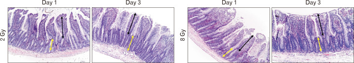
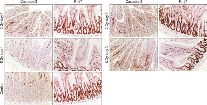
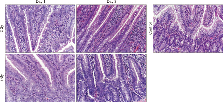
Morphological evaluation of the small intestine mucosa and apoptosis activity (caspase-3) is necessary to assess the severity of damage to the small intestine. At the same time, proliferative index based on Ki-67 can be used to assess the regenerative potential of the small intestine. Fragments of small intestine of Wistar rats (n=60) of three groups: I) control (n=20); II) experimental group (n=20; local single electron irradiation at a dose of 2 Gy), III) experimental group (n=20; local single electron irradiation at a dose of 8 Gy) were studied by light microscopy using hematoxylin and eosin staining and immunohistochemical reactions with antibodies to Ki-67 and caspase-3. In all samples of the experimental groups, a decrease in all morphometric indices was observed on day 1 with a tendency to recover on day 3. Small intestinal electron irradiation led to disturbances in the histoarchitecture of varying severity, and an increase in cell apoptosis was observed (increased expression of caspase-3 and decrease in Ki-67). In addition, modulation of the PI3K/AKT and MAPK/ERK signaling pathways was detected. The most pronounced destructive changes were observed in the group of 8 Gy single electron irradiation. Local irradiation of the small intestine with electrons at a dose of 2 and 8 Gy results in a decrease in the number of enterocytes, mainly stem cells of the intestinal crypts.


 分享
分享
 求助内容:
求助内容: 应助结果提醒方式:
 扫码关注我们
