{"title":"Colonic black spots and patches in a 50-year-old woman","authors":"Puo-Hsien Le, Tse-Ching Chen, Cheng-Tang Chiu","doi":"10.1002/aid2.13405","DOIUrl":null,"url":null,"abstract":"<p>This 50-year-old woman had no systemic disease. She received health examination, and colonoscopy found multiple back spots and patches from cecum to descending colon, especially proximal colon (Figure 1A,B). Colonic biopsies were obtained from pigmented lesions, and a representative hematoxylin and eosin stain (Figure 1C) and a Fontana-Masson stain (Figure 1D) were shown. No similar discoloration was noted in upper alimentary tract to the duodenum by endoscopy. Histology of the discolored colonic lesion showed colonic mucosa with scattered nests of melanocytic-like cells in mucosa and submucosa (Figure 1C). The brown pigment was positive for Fontana-Masson stain (Figure 1D) and negative for iron and PAS stain. Therefore, it was melanin. In immunohistochemical study, the pigmented cells were positive for HMB-45, S-100, and Melan-A expression, negative for CD163 expression and Ki-67/MIB-1 labeling index labeling index <2%. Therefore, the diagnosis was melanocytic nevus.</p><p>Typical melanocytic nevi are round with a uniform color and a diameter of 5 mm or less on the skin.<span><sup>1</sup></span> It is caused by proliferation of melanocytes, and associated with ~30% of melanomas.<span><sup>2</sup></span> Colonic melanocytic nevi are also regarded as potential precursor lesions of malignant melanoma.<span><sup>3</sup></span> Only one case of colonic melanocytic nevi with completely pathological diagnosis has been reported.<span><sup>4</sup></span> In that case, the lesion is a single brownish flat area occupying a quarter of the colonic wall in the ascending colon. However, we presented the case with diffuse black spots and patches on colonic mucosa.</p><p>Unlike melanosis coli, which shows continuous homogeneous black-brownish discoloration of colon mucosa (snake-skin appearance or starry sky appearance), melanosis nevus has round pigmentations with heterogenous distribution.<span><sup>5</sup></span> Microscopically, melanosis coli is characterized by deposition of lipofuscin in histiocytes, while melanocytic proliferation is noted in the melanocytic nevus of colon.</p><p>Puo-Hsien Le performed the colonoscopy and drafted the article. Tse-Ching Chen confirmed the diagnosis. Cheng-Tang Chiu revised the article critically for important intellectual content. All authors had final approval of the version to be submitted.</p><p>The authors declare no conflicts of interest.</p><p>Yes.</p>","PeriodicalId":7278,"journal":{"name":"Advances in Digestive Medicine","volume":"11 4","pages":"228-229"},"PeriodicalIF":0.4000,"publicationDate":"2024-06-05","publicationTypes":"Journal Article","fieldsOfStudy":null,"isOpenAccess":false,"openAccessPdf":"https://onlinelibrary.wiley.com/doi/epdf/10.1002/aid2.13405","citationCount":"0","resultStr":null,"platform":"Semanticscholar","paperid":null,"PeriodicalName":"Advances in Digestive Medicine","FirstCategoryId":"1085","ListUrlMain":"https://onlinelibrary.wiley.com/doi/10.1002/aid2.13405","RegionNum":0,"RegionCategory":null,"ArticlePicture":[],"TitleCN":null,"AbstractTextCN":null,"PMCID":null,"EPubDate":"","PubModel":"","JCR":"Q4","JCRName":"GASTROENTEROLOGY & HEPATOLOGY","Score":null,"Total":0}
引用次数: 0
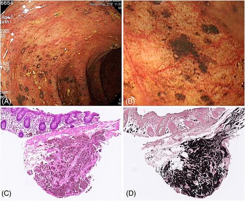
Abstract
This 50-year-old woman had no systemic disease. She received health examination, and colonoscopy found multiple back spots and patches from cecum to descending colon, especially proximal colon (Figure 1A,B). Colonic biopsies were obtained from pigmented lesions, and a representative hematoxylin and eosin stain (Figure 1C) and a Fontana-Masson stain (Figure 1D) were shown. No similar discoloration was noted in upper alimentary tract to the duodenum by endoscopy. Histology of the discolored colonic lesion showed colonic mucosa with scattered nests of melanocytic-like cells in mucosa and submucosa (Figure 1C). The brown pigment was positive for Fontana-Masson stain (Figure 1D) and negative for iron and PAS stain. Therefore, it was melanin. In immunohistochemical study, the pigmented cells were positive for HMB-45, S-100, and Melan-A expression, negative for CD163 expression and Ki-67/MIB-1 labeling index labeling index <2%. Therefore, the diagnosis was melanocytic nevus.
Typical melanocytic nevi are round with a uniform color and a diameter of 5 mm or less on the skin.1 It is caused by proliferation of melanocytes, and associated with ~30% of melanomas.2 Colonic melanocytic nevi are also regarded as potential precursor lesions of malignant melanoma.3 Only one case of colonic melanocytic nevi with completely pathological diagnosis has been reported.4 In that case, the lesion is a single brownish flat area occupying a quarter of the colonic wall in the ascending colon. However, we presented the case with diffuse black spots and patches on colonic mucosa.
Unlike melanosis coli, which shows continuous homogeneous black-brownish discoloration of colon mucosa (snake-skin appearance or starry sky appearance), melanosis nevus has round pigmentations with heterogenous distribution.5 Microscopically, melanosis coli is characterized by deposition of lipofuscin in histiocytes, while melanocytic proliferation is noted in the melanocytic nevus of colon.
Puo-Hsien Le performed the colonoscopy and drafted the article. Tse-Ching Chen confirmed the diagnosis. Cheng-Tang Chiu revised the article critically for important intellectual content. All authors had final approval of the version to be submitted.
期刊介绍:
Advances in Digestive Medicine is the official peer-reviewed journal of GEST, DEST and TASL. Missions of AIDM are to enhance the quality of patient care, to promote researches in gastroenterology, endoscopy and hepatology related fields, and to develop platforms for digestive science. Specific areas of interest are included, but not limited to: • Acid-related disease • Small intestinal disease • Digestive cancer • Diagnostic & therapeutic endoscopy • Enteral nutrition • Innovation in endoscopic technology • Functional GI • Hepatitis • GI images • Liver cirrhosis • Gut hormone • NASH • Helicobacter pylori • Cancer screening • IBD • Laparoscopic surgery • Infectious disease of digestive tract • Genetics and metabolic disorder • Microbiota • Regenerative medicine • Pancreaticobiliary disease • Guideline & consensus.


 分享
分享
 求助内容:
求助内容: 应助结果提醒方式:
 扫码关注我们
