Marianna Angelo Palmejani Albacete, Gustavo Novelino Simão, Charles Marques Lourenço, Antonio Carlos Dos Santos
{"title":"Vanishing white matter disease: imaging, clinical and molecular correlation in Brazilian families.","authors":"Marianna Angelo Palmejani Albacete, Gustavo Novelino Simão, Charles Marques Lourenço, Antonio Carlos Dos Santos","doi":"10.1007/s00234-024-03405-z","DOIUrl":null,"url":null,"abstract":"<p><strong>Purpose: </strong>To characterize Vanishing White Matter Disease (VWM) cases from a Brazilian University Tertiary hospital, focusing on brain magnetic resonance image (MRI) aspects, clinical and molecular data.</p><p><strong>Methods: </strong>Medical records and brain MRI of 13 genetically confirmed VWM patients were reviewed. Epidemiological data such as age at symptom onset, gender and main symptoms were analyzed, along with genetic mutations and MRI characteristics, such as the distribution of white matter lesions and atrophy.</p><p><strong>Results: </strong>The majority of patients were female, with the age of symptom onset ranging from 1 year and 6 months to 40 years. All mutations were identified in the EIF2B5 gene, the most prevalent being c.338G > A (p.Arg113His), and a novel mutation related to the disease was discovered, c.1051G > A (p.Gly351Ser). Trauma or infection were significant triggers. The most frequent symptoms were ataxia and limb spasticity. All MRI scans displayed deep white matter involvement, cystic degeneration, with U-fibers relatively spared and a predilection for the frontoparietal region. Lesions in the corpus callosum and posterior fossa were present in all patients. Follow-up exams revealed the evolution of white matter lesions and cerebral atrophy, which correlated with clinical deterioration.</p><p><strong>Conclusions: </strong>VWM affects various age groups, with a significant clinical and genetic variability. A novel mutation associated with the disease is highlighted. MRI reveals a typical pattern of white matter involvement, characterized by diffuse lesions in the periventricular and deep regions, with subsequent extension to the subcortical areas, accompanied by cystic degeneration, and plays a crucial role in diagnosis and follow-up.</p>","PeriodicalId":19422,"journal":{"name":"Neuroradiology","volume":" ","pages":"1553-1564"},"PeriodicalIF":2.6000,"publicationDate":"2024-09-01","publicationTypes":"Journal Article","fieldsOfStudy":null,"isOpenAccess":false,"openAccessPdf":"","citationCount":"0","resultStr":null,"platform":"Semanticscholar","paperid":null,"PeriodicalName":"Neuroradiology","FirstCategoryId":"3","ListUrlMain":"https://doi.org/10.1007/s00234-024-03405-z","RegionNum":3,"RegionCategory":"医学","ArticlePicture":[],"TitleCN":null,"AbstractTextCN":null,"PMCID":null,"EPubDate":"2024/6/18 0:00:00","PubModel":"Epub","JCR":"Q2","JCRName":"CLINICAL NEUROLOGY","Score":null,"Total":0}
引用次数: 0
Abstract
Purpose: To characterize Vanishing White Matter Disease (VWM) cases from a Brazilian University Tertiary hospital, focusing on brain magnetic resonance image (MRI) aspects, clinical and molecular data.
Methods: Medical records and brain MRI of 13 genetically confirmed VWM patients were reviewed. Epidemiological data such as age at symptom onset, gender and main symptoms were analyzed, along with genetic mutations and MRI characteristics, such as the distribution of white matter lesions and atrophy.
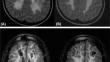
Results: The majority of patients were female, with the age of symptom onset ranging from 1 year and 6 months to 40 years. All mutations were identified in the EIF2B5 gene, the most prevalent being c.338G > A (p.Arg113His), and a novel mutation related to the disease was discovered, c.1051G > A (p.Gly351Ser). Trauma or infection were significant triggers. The most frequent symptoms were ataxia and limb spasticity. All MRI scans displayed deep white matter involvement, cystic degeneration, with U-fibers relatively spared and a predilection for the frontoparietal region. Lesions in the corpus callosum and posterior fossa were present in all patients. Follow-up exams revealed the evolution of white matter lesions and cerebral atrophy, which correlated with clinical deterioration.
Conclusions: VWM affects various age groups, with a significant clinical and genetic variability. A novel mutation associated with the disease is highlighted. MRI reveals a typical pattern of white matter involvement, characterized by diffuse lesions in the periventricular and deep regions, with subsequent extension to the subcortical areas, accompanied by cystic degeneration, and plays a crucial role in diagnosis and follow-up.
期刊介绍:
Neuroradiology aims to provide state-of-the-art medical and scientific information in the fields of Neuroradiology, Neurosciences, Neurology, Psychiatry, Neurosurgery, and related medical specialities. Neuroradiology as the official Journal of the European Society of Neuroradiology receives submissions from all parts of the world and publishes peer-reviewed original research, comprehensive reviews, educational papers, opinion papers, and short reports on exceptional clinical observations and new technical developments in the field of Neuroimaging and Neurointervention. The journal has subsections for Diagnostic and Interventional Neuroradiology, Advanced Neuroimaging, Paediatric Neuroradiology, Head-Neck-ENT Radiology, Spine Neuroradiology, and for submissions from Japan. Neuroradiology aims to provide new knowledge about and insights into the function and pathology of the human nervous system that may help to better diagnose and treat nervous system diseases. Neuroradiology is a member of the Committee on Publication Ethics (COPE) and follows the COPE core practices. Neuroradiology prefers articles that are free of bias, self-critical regarding limitations, transparent and clear in describing study participants, methods, and statistics, and short in presenting results. Before peer-review all submissions are automatically checked by iThenticate to assess for potential overlap in prior publication.


 分享
分享
 求助内容:
求助内容: 应助结果提醒方式:
 扫码关注我们
