A pretreatment multiparametric MRI-based radiomics-clinical machine learning model for predicting radiation-induced temporal lobe injury in patients with nasopharyngeal carcinoma
Li Wang MS, Ting Qiu MS, Jiawei Zhou MS, Yinsu Zhu MD, PhD, Baozhou Sun PhD, Guanyu Yang PhD, Shengfu Huang MD, Lirong Wu MD, PhD, Xia He MD, PhD
{"title":"A pretreatment multiparametric MRI-based radiomics-clinical machine learning model for predicting radiation-induced temporal lobe injury in patients with nasopharyngeal carcinoma","authors":"Li Wang MS, Ting Qiu MS, Jiawei Zhou MS, Yinsu Zhu MD, PhD, Baozhou Sun PhD, Guanyu Yang PhD, Shengfu Huang MD, Lirong Wu MD, PhD, Xia He MD, PhD","doi":"10.1002/hed.27830","DOIUrl":null,"url":null,"abstract":"<div>\n \n \n <section>\n \n <h3> Background</h3>\n \n <p>To establish and validate a machine learning model using pretreatment multiparametric magnetic resonance imaging-based radiomics data with clinical data to predict radiation-induced temporal lobe injury (RTLI) in patients with nasopharyngeal carcinoma (NPC) after intensity-modulated radiotherapy (IMRT).</p>\n </section>\n \n <section>\n \n <h3> Methods</h3>\n \n <p>Data from 230 patients with NPC who received IMRT (130 with RTLI and 130 without) were randomly divided into the training (<i>n</i> = 161) and validation cohort (<i>n</i> = 69) with a ratio of 7:3. Radiomics features were extracted from pretreatment apparent diffusion coefficient (ADC) map, T2-weighted imaging (T2WI), and CE-T1-weighted imaging (CE-T1WI). <i>T</i>-test, spearman rank correlation, and least absolute shrinkage and selection operator (LASSO) algorithm were employed to identify significant radiomics features. Clinical features were selected with univariate and multivariate analyses. Radiomics and clinical models were constructed using multiple machine learning classifiers, and a clinical-radiomics nomogram that combined clinical with radiomics features was developed. Receiver operating characteristic (ROC) curves, calibration curves, and decision curve analysis (DCA) were drawn to compare and verify the predictive performances of the clinical model, radiomics model, and clinical-radiomics nomogram.</p>\n </section>\n \n <section>\n \n <h3> Results</h3>\n \n <p>A total of 5064 radiomics features were extracted, from which 52 radiomics features were selected to construct the radiomics signature. The AUC of the radiomics signature based on multiparametric MRI was 0.980 in the training cohort and 0.969 in the validation cohort, outperforming the radiomics signature only based on T2WI and CE-T1WI (<i>p</i> < 0.05), which highlighted the significance of the DWI sequence in the prediction of temporal lobe injury. The area under the curve (AUC) of the clinical model was 0.895 in the training cohort and 0.905 in the validation cohort. The nomogram, which integrated radiomics and clinical features, demonstrated an impressive AUC value of 0.984 in the validation set; however, no statistically significant difference was observed compared to the radiomics model. The calibration curve and decision curve analysis of the nomogram demonstrated excellent predictive performance and clinical feasibility.</p>\n </section>\n \n <section>\n \n <h3> Conclusions</h3>\n \n <p>The clinical-radiomics nomogram, integrating clinical features with radiomics features derived from pretreatment multiparametric MRI, exhibits compelling predictive performance for RTLI in patients diagnosed with NPC.</p>\n </section>\n </div>","PeriodicalId":55072,"journal":{"name":"Head and Neck-Journal for the Sciences and Specialties of the Head and Neck","volume":"46 9","pages":"2132-2144"},"PeriodicalIF":2.2000,"publicationDate":"2024-06-18","publicationTypes":"Journal Article","fieldsOfStudy":null,"isOpenAccess":false,"openAccessPdf":"https://onlinelibrary.wiley.com/doi/epdf/10.1002/hed.27830","citationCount":"0","resultStr":null,"platform":"Semanticscholar","paperid":null,"PeriodicalName":"Head and Neck-Journal for the Sciences and Specialties of the Head and Neck","FirstCategoryId":"3","ListUrlMain":"https://onlinelibrary.wiley.com/doi/10.1002/hed.27830","RegionNum":3,"RegionCategory":"医学","ArticlePicture":[],"TitleCN":null,"AbstractTextCN":null,"PMCID":null,"EPubDate":"","PubModel":"","JCR":"Q1","JCRName":"OTORHINOLARYNGOLOGY","Score":null,"Total":0}
引用次数: 0
Abstract
Background
To establish and validate a machine learning model using pretreatment multiparametric magnetic resonance imaging-based radiomics data with clinical data to predict radiation-induced temporal lobe injury (RTLI) in patients with nasopharyngeal carcinoma (NPC) after intensity-modulated radiotherapy (IMRT).
Methods
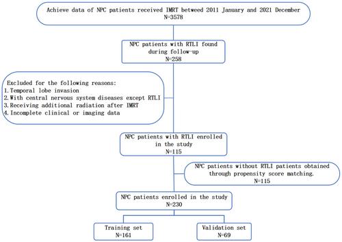
Data from 230 patients with NPC who received IMRT (130 with RTLI and 130 without) were randomly divided into the training (n = 161) and validation cohort (n = 69) with a ratio of 7:3. Radiomics features were extracted from pretreatment apparent diffusion coefficient (ADC) map, T2-weighted imaging (T2WI), and CE-T1-weighted imaging (CE-T1WI). T-test, spearman rank correlation, and least absolute shrinkage and selection operator (LASSO) algorithm were employed to identify significant radiomics features. Clinical features were selected with univariate and multivariate analyses. Radiomics and clinical models were constructed using multiple machine learning classifiers, and a clinical-radiomics nomogram that combined clinical with radiomics features was developed. Receiver operating characteristic (ROC) curves, calibration curves, and decision curve analysis (DCA) were drawn to compare and verify the predictive performances of the clinical model, radiomics model, and clinical-radiomics nomogram.
Results
A total of 5064 radiomics features were extracted, from which 52 radiomics features were selected to construct the radiomics signature. The AUC of the radiomics signature based on multiparametric MRI was 0.980 in the training cohort and 0.969 in the validation cohort, outperforming the radiomics signature only based on T2WI and CE-T1WI (p < 0.05), which highlighted the significance of the DWI sequence in the prediction of temporal lobe injury. The area under the curve (AUC) of the clinical model was 0.895 in the training cohort and 0.905 in the validation cohort. The nomogram, which integrated radiomics and clinical features, demonstrated an impressive AUC value of 0.984 in the validation set; however, no statistically significant difference was observed compared to the radiomics model. The calibration curve and decision curve analysis of the nomogram demonstrated excellent predictive performance and clinical feasibility.
Conclusions
The clinical-radiomics nomogram, integrating clinical features with radiomics features derived from pretreatment multiparametric MRI, exhibits compelling predictive performance for RTLI in patients diagnosed with NPC.
期刊介绍:
Head & Neck is an international multidisciplinary publication of original contributions concerning the diagnosis and management of diseases of the head and neck. This area involves the overlapping interests and expertise of several surgical and medical specialties, including general surgery, neurosurgery, otolaryngology, plastic surgery, oral surgery, dermatology, ophthalmology, pathology, radiotherapy, medical oncology, and the corresponding basic sciences.


 分享
分享
 求助内容:
求助内容: 应助结果提醒方式:
 扫码关注我们
