Ilhem Lilia Jaabar, Brittany Foley, Alberto Mezzetti, Françoise Pillier, Francis Berenbaum, Jessem Landoulsi, Xavier Houard
{"title":"Unraveling the Mechanisms of Hypertrophy-Induced Matrix Mineralization and Modifications in Articular Chondrocytes.","authors":"Ilhem Lilia Jaabar, Brittany Foley, Alberto Mezzetti, Françoise Pillier, Francis Berenbaum, Jessem Landoulsi, Xavier Houard","doi":"10.1007/s00223-024-01229-w","DOIUrl":null,"url":null,"abstract":"<p><p>Chondrocyte hypertrophic differentiation is a main event leading to articular cartilage degradation in osteoarthritis. It is associated with matrix remodeling and mineralization, the dynamics of which is not well characterized during chondrocyte hypertrophic differentiation in articular cartilage. Based on an in vitro model of progressive differentiation of immature murine articular chondrocytes (iMACs) into prehypertrophic (Prehyp) and hypertrophic (Hyp) chondrocytes, we performed kinetics of chondrocyte differentiation from Prehyp to Hyp to follow matrix mineralization and remodeling by immunofluorescence, biochemical, molecular, and physicochemical approaches, including atomic force microscopy, scanning electron microscopy associated with energy-dispersive X-ray spectroscopy (SEM-EDS), attenuated total reflection infrared analyses, and X-ray diffraction. Chondrocyte apoptosis was determined by TUNEL assay. The results show the formation of a mineral phase 7 days after Hyp induction, which spreads within the matrices to form poorly crystalline carbonate-substituted hydroxyapatite after 14 days, then the proportions of crystalline relative to amorphous content increases over time. Hyp differentiation also induced a matrix turnover that occurs over the first 7 days, characterized by a decrease in type II collagen and aggrecan and the concomitant appearance of type X collagen. This is accompanied by an increase in the enzymatic activity of MMP-13, the main collagenase in cartilage. The number of apoptotic chondrocytes slightly increased with Hyp differentiation and SEM-EDS analyses detected phosphorus-rich structures that could correspond to apoptotic bodies. Our findings highlight the mechanisms of matrix remodeling events leading to the mineralization of articular cartilage that may occur in osteoarthritis.</p>","PeriodicalId":9601,"journal":{"name":"Calcified Tissue International","volume":" ","pages":"269-282"},"PeriodicalIF":3.2000,"publicationDate":"2024-09-01","publicationTypes":"Journal Article","fieldsOfStudy":null,"isOpenAccess":false,"openAccessPdf":"","citationCount":"0","resultStr":null,"platform":"Semanticscholar","paperid":null,"PeriodicalName":"Calcified Tissue International","FirstCategoryId":"3","ListUrlMain":"https://doi.org/10.1007/s00223-024-01229-w","RegionNum":3,"RegionCategory":"医学","ArticlePicture":[],"TitleCN":null,"AbstractTextCN":null,"PMCID":null,"EPubDate":"2024/6/25 0:00:00","PubModel":"Epub","JCR":"Q2","JCRName":"ENDOCRINOLOGY & METABOLISM","Score":null,"Total":0}
引用次数: 0
Abstract
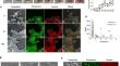
Chondrocyte hypertrophic differentiation is a main event leading to articular cartilage degradation in osteoarthritis. It is associated with matrix remodeling and mineralization, the dynamics of which is not well characterized during chondrocyte hypertrophic differentiation in articular cartilage. Based on an in vitro model of progressive differentiation of immature murine articular chondrocytes (iMACs) into prehypertrophic (Prehyp) and hypertrophic (Hyp) chondrocytes, we performed kinetics of chondrocyte differentiation from Prehyp to Hyp to follow matrix mineralization and remodeling by immunofluorescence, biochemical, molecular, and physicochemical approaches, including atomic force microscopy, scanning electron microscopy associated with energy-dispersive X-ray spectroscopy (SEM-EDS), attenuated total reflection infrared analyses, and X-ray diffraction. Chondrocyte apoptosis was determined by TUNEL assay. The results show the formation of a mineral phase 7 days after Hyp induction, which spreads within the matrices to form poorly crystalline carbonate-substituted hydroxyapatite after 14 days, then the proportions of crystalline relative to amorphous content increases over time. Hyp differentiation also induced a matrix turnover that occurs over the first 7 days, characterized by a decrease in type II collagen and aggrecan and the concomitant appearance of type X collagen. This is accompanied by an increase in the enzymatic activity of MMP-13, the main collagenase in cartilage. The number of apoptotic chondrocytes slightly increased with Hyp differentiation and SEM-EDS analyses detected phosphorus-rich structures that could correspond to apoptotic bodies. Our findings highlight the mechanisms of matrix remodeling events leading to the mineralization of articular cartilage that may occur in osteoarthritis.
软骨细胞肥大分化是导致骨关节炎关节软骨退化的主要原因。它与基质重塑和矿化有关,而关节软骨中软骨细胞肥大分化过程的动态特征尚不清楚。基于未成熟小鼠关节软骨细胞(iMACs)逐渐分化为肥大前(Prehyp)和肥大(Hyp)软骨细胞的体外模型,我们进行了软骨细胞从Prehyp到Hyp分化的动力学研究,并通过免疫荧光跟踪基质矿化和重塑、生化、分子和物理化学方法,包括原子力显微镜、扫描电子显微镜与能量色散 X 射线光谱(SEM-EDS)、衰减全反射红外分析和 X 射线衍射。软骨细胞凋亡通过 TUNEL 检测法进行测定。结果表明,Hyp诱导7天后形成矿物相,14天后矿物相在基质内扩散,形成结晶度较差的碳酸盐取代羟基磷灰石,然后随着时间的推移,结晶相对于无定形含量的比例增加。Hyp 分化还诱导基质在最初 7 天内发生更替,其特点是 II 型胶原蛋白和凝集素减少,同时出现 X 型胶原蛋白。与此同时,软骨中主要的胶原酶 MMP-13 的酶活性增加。随着 Hyp 分化,凋亡软骨细胞的数量略有增加,SEM-EDS 分析检测到富含磷的结构,可能与凋亡体相对应。我们的研究结果强调了骨关节炎中可能发生的导致关节软骨矿化的基质重塑事件的机制。
期刊介绍:
Calcified Tissue International and Musculoskeletal Research publishes original research and reviews concerning the structure and function of bone, and other musculoskeletal tissues in living organisms and clinical studies of musculoskeletal disease. It includes studies of cell biology, molecular biology, intracellular signalling, and physiology, as well as research into the hormones, cytokines and other mediators that influence the musculoskeletal system. The journal also publishes clinical studies of relevance to bone disease, mineral metabolism, muscle function, and musculoskeletal interactions.


 分享
分享
 求助内容:
求助内容: 应助结果提醒方式:
 扫码关注我们
