Virgilio Mb Braga, Ana Gs Limoeiro, Marilia Fv Marceliano-Alves, Marcelo Coelho, Alessandra Machado, Ricardo T Lopes, Wayne M Nascimento, Adriana J Soares, Marcos Frozoni
{"title":"Efficacy of WaveOne Gold and ProDesign RT systems in removing filling material: a micro-CT analysis.","authors":"Virgilio Mb Braga, Ana Gs Limoeiro, Marilia Fv Marceliano-Alves, Marcelo Coelho, Alessandra Machado, Ricardo T Lopes, Wayne M Nascimento, Adriana J Soares, Marcos Frozoni","doi":"10.54589/aol.37/1/34","DOIUrl":null,"url":null,"abstract":"<p><p>The remaining filling material after retreatment can harbor bacteria and organic tissues that can influence the outcome of the therapy.</p><p><strong>Aim: </strong>The aim of this study was to evalúate, by micro-CT, the amount of filling material remaining in the root canal after its removal using WaveOne Gold or ProDesign RT.</p><p><strong>Material and method: </strong>Forty human mandibular canines were instrumented with the ProTaper Next system up to the X2 instrument (25.06) and filled with gutta-percha cones and AHPlus. Teeth were divided into 2 groups (n=20): WaveOne Gold 25.07 (WOG) and ProDesign RT 25.08 (PRT) for filling removal, after which they were scanned in a micro-CT device to quantify the volume of remaining filling material. The data were subjected to log <sup>10</sup> transformation, Student 's t-test was performed to account for multiple observationsper sample, significance was set at 5%.</p><p><strong>Results: </strong>Student 's t-test showed that there was no difference between the two systems regarding the volume of remaining filling material in the thirds: apical (p = 0.392), middle (p = 0.065), or cervical (p = 0.918).</p><p><strong>Conclusión: </strong>Remaining filling material was present in all groups and both systems were similar in removing root filling material in mandibular canines.</p>","PeriodicalId":93853,"journal":{"name":"Acta odontologica latinoamericana : AOL","volume":"37 1","pages":"34-39"},"PeriodicalIF":0.0000,"publicationDate":"2024-04-01","publicationTypes":"Journal Article","fieldsOfStudy":null,"isOpenAccess":false,"openAccessPdf":"https://www.ncbi.nlm.nih.gov/pmc/articles/PMC11212326/pdf/","citationCount":"0","resultStr":null,"platform":"Semanticscholar","paperid":null,"PeriodicalName":"Acta odontologica latinoamericana : AOL","FirstCategoryId":"1085","ListUrlMain":"https://doi.org/10.54589/aol.37/1/34","RegionNum":0,"RegionCategory":null,"ArticlePicture":[],"TitleCN":null,"AbstractTextCN":null,"PMCID":null,"EPubDate":"","PubModel":"","JCR":"","JCRName":"","Score":null,"Total":0}
引用次数: 0
Abstract
The remaining filling material after retreatment can harbor bacteria and organic tissues that can influence the outcome of the therapy.
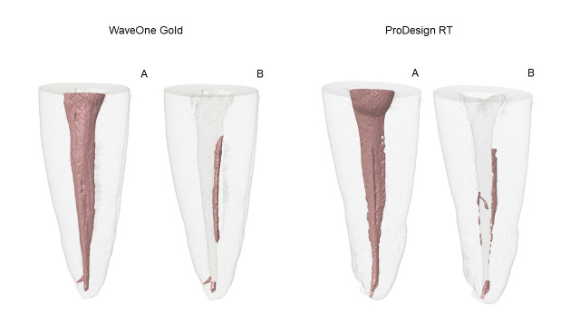
Aim: The aim of this study was to evalúate, by micro-CT, the amount of filling material remaining in the root canal after its removal using WaveOne Gold or ProDesign RT.
Material and method: Forty human mandibular canines were instrumented with the ProTaper Next system up to the X2 instrument (25.06) and filled with gutta-percha cones and AHPlus. Teeth were divided into 2 groups (n=20): WaveOne Gold 25.07 (WOG) and ProDesign RT 25.08 (PRT) for filling removal, after which they were scanned in a micro-CT device to quantify the volume of remaining filling material. The data were subjected to log 10 transformation, Student 's t-test was performed to account for multiple observationsper sample, significance was set at 5%.
Results: Student 's t-test showed that there was no difference between the two systems regarding the volume of remaining filling material in the thirds: apical (p = 0.392), middle (p = 0.065), or cervical (p = 0.918).
Conclusión: Remaining filling material was present in all groups and both systems were similar in removing root filling material in mandibular canines.


 分享
分享

 求助内容:
求助内容: 应助结果提醒方式:
 扫码关注我们
