Alexander Rau, Gabriel Gonzalez-Escamilla, Nils Schroeter, Ahmed Othman, Andrea Dressing, Cornelius Weiller, Horst Urbach, Marco Reisert, Sergiu Groppa, Jonas A. Hosp
{"title":"Inflammation-Triggered Enlargement of Choroid Plexus in Subacute COVID-19 Patients with Neurological Symptoms","authors":"Alexander Rau, Gabriel Gonzalez-Escamilla, Nils Schroeter, Ahmed Othman, Andrea Dressing, Cornelius Weiller, Horst Urbach, Marco Reisert, Sergiu Groppa, Jonas A. Hosp","doi":"10.1002/ana.27016","DOIUrl":null,"url":null,"abstract":"<div>\n \n <section>\n \n <h3> Objective</h3>\n \n <p>To investigate whether choroid plexus volumes in subacute coronavirus disease 2019 (COVID-19) patients with neurological symptoms could indicate inflammatory activation or barrier dysfunction and assess their association with clinical data.</p>\n </section>\n \n <section>\n \n <h3> Methods</h3>\n \n <p>Choroid plexus volumes were measured in 28 subacute COVID-19 patients via cerebral magnetic resonance imaging (MRI), compared with those in infection-triggered non-COVID-19 encephalopathy patients (n = 25), asymptomatic individuals after COVID-19 (n = 21), and healthy controls (n = 21). Associations with inflammatory serum markers (peak counts of leukocytes, C-reactive protein [CRP], interleukin 6), an MRI-based marker of barrier dysfunction (CSF volume fraction [V-CSF]), and clinical parameters like olfactory performance and cognitive scores (Montreal Cognitive Assessment) were investigated.</p>\n </section>\n \n <section>\n \n <h3> Results</h3>\n \n <p>COVID-19 patients showed significantly larger choroid plexus volumes than control groups (<i>p</i> < 0.001, η<sup>2</sup> = 0.172). These volumes correlated significantly with peak leukocyte levels (<i>p</i> = 0.001, Pearson's r = 0.621) and V-CSF (<i>p</i> = 0.009, Spearman's rho = 0.534), but neither with CRP nor interleukin 6. No significant correlations were found with clinical parameters.</p>\n </section>\n \n <section>\n \n <h3> Interpretation</h3>\n \n <p>In patients with subacute COVID-19, choroid plexus volume is a marker of central nervous system inflammation and barrier dysfunction in the presence of neurologic symptoms. The absence of plexus enlargement in infection-triggered non-COVID-19 encephalopathy suggests a specific severe acute respiratory syndrome coronavirus 2 effect. This study also documents an increase in choroid plexus volume for the first time as a parainfectious event. ANN NEUROL 2024;96:715–725</p>\n </section>\n </div>","PeriodicalId":127,"journal":{"name":"Annals of Neurology","volume":"96 4","pages":"715-725"},"PeriodicalIF":7.7000,"publicationDate":"2024-06-27","publicationTypes":"Journal Article","fieldsOfStudy":null,"isOpenAccess":false,"openAccessPdf":"https://onlinelibrary.wiley.com/doi/epdf/10.1002/ana.27016","citationCount":"0","resultStr":null,"platform":"Semanticscholar","paperid":null,"PeriodicalName":"Annals of Neurology","FirstCategoryId":"3","ListUrlMain":"https://onlinelibrary.wiley.com/doi/10.1002/ana.27016","RegionNum":1,"RegionCategory":"医学","ArticlePicture":[],"TitleCN":null,"AbstractTextCN":null,"PMCID":null,"EPubDate":"","PubModel":"","JCR":"Q1","JCRName":"CLINICAL NEUROLOGY","Score":null,"Total":0}
引用次数: 0
Abstract
Objective
To investigate whether choroid plexus volumes in subacute coronavirus disease 2019 (COVID-19) patients with neurological symptoms could indicate inflammatory activation or barrier dysfunction and assess their association with clinical data.
Methods
Choroid plexus volumes were measured in 28 subacute COVID-19 patients via cerebral magnetic resonance imaging (MRI), compared with those in infection-triggered non-COVID-19 encephalopathy patients (n = 25), asymptomatic individuals after COVID-19 (n = 21), and healthy controls (n = 21). Associations with inflammatory serum markers (peak counts of leukocytes, C-reactive protein [CRP], interleukin 6), an MRI-based marker of barrier dysfunction (CSF volume fraction [V-CSF]), and clinical parameters like olfactory performance and cognitive scores (Montreal Cognitive Assessment) were investigated.
Results
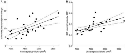
COVID-19 patients showed significantly larger choroid plexus volumes than control groups (p < 0.001, η2 = 0.172). These volumes correlated significantly with peak leukocyte levels (p = 0.001, Pearson's r = 0.621) and V-CSF (p = 0.009, Spearman's rho = 0.534), but neither with CRP nor interleukin 6. No significant correlations were found with clinical parameters.
Interpretation
In patients with subacute COVID-19, choroid plexus volume is a marker of central nervous system inflammation and barrier dysfunction in the presence of neurologic symptoms. The absence of plexus enlargement in infection-triggered non-COVID-19 encephalopathy suggests a specific severe acute respiratory syndrome coronavirus 2 effect. This study also documents an increase in choroid plexus volume for the first time as a parainfectious event. ANN NEUROL 2024;96:715–725
期刊介绍:
Annals of Neurology publishes original articles with potential for high impact in understanding the pathogenesis, clinical and laboratory features, diagnosis, treatment, outcomes and science underlying diseases of the human nervous system. Articles should ideally be of broad interest to the academic neurological community rather than solely to subspecialists in a particular field. Studies involving experimental model system, including those in cell and organ cultures and animals, of direct translational relevance to the understanding of neurological disease are also encouraged.


 分享
分享
 求助内容:
求助内容: 应助结果提醒方式:
 扫码关注我们
