{"title":"Ultrasonographic diagnosis of cervical lymph nodes.","authors":"Daisuke Saito, Kiyoto Shiga","doi":"10.1007/s10396-024-01466-4","DOIUrl":null,"url":null,"abstract":"<p><p>Many patients visit outpatient clinics suffering from cervical lymphadenopathy. For those patients, ultrasonography is useful in differentiating inflammatory diseases and malignant tumors. On ultrasonographic images, normal lymph nodes are indicated as hypoechogenic masses with a well-defined border. The medullary portion near the lymph node hilum is hyperechogenic, so-called fatty hilum (FH). Color Doppler imaging reveals that blood flows from the lymph node hilum to FH. In lymph node metastasis, a metastatic focus grows within lymph nodes, which displaces and destroys the structure of normal lymph nodes. Ultrasonography can be used to detect FH, disappearance and unevenness of blood flow within lymph nodes, cyst formation, and so on. It is important to closely observe the inside of lymph nodes and make a diagnosis via ultrasonography, based on the criteria for diagnosing lymph node metastasis from head and neck squamous cell carcinoma. Additionally, it is also necessary to distinguish among inflammatory lymphadenopathy and malignant lymphoma.</p>","PeriodicalId":50130,"journal":{"name":"Journal of Medical Ultrasonics","volume":" ","pages":""},"PeriodicalIF":2.1000,"publicationDate":"2024-06-28","publicationTypes":"Journal Article","fieldsOfStudy":null,"isOpenAccess":false,"openAccessPdf":"","citationCount":"0","resultStr":null,"platform":"Semanticscholar","paperid":null,"PeriodicalName":"Journal of Medical Ultrasonics","FirstCategoryId":"3","ListUrlMain":"https://doi.org/10.1007/s10396-024-01466-4","RegionNum":4,"RegionCategory":"医学","ArticlePicture":[],"TitleCN":null,"AbstractTextCN":null,"PMCID":null,"EPubDate":"","PubModel":"","JCR":"Q3","JCRName":"RADIOLOGY, NUCLEAR MEDICINE & MEDICAL IMAGING","Score":null,"Total":0}
引用次数: 0
Abstract
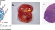
Many patients visit outpatient clinics suffering from cervical lymphadenopathy. For those patients, ultrasonography is useful in differentiating inflammatory diseases and malignant tumors. On ultrasonographic images, normal lymph nodes are indicated as hypoechogenic masses with a well-defined border. The medullary portion near the lymph node hilum is hyperechogenic, so-called fatty hilum (FH). Color Doppler imaging reveals that blood flows from the lymph node hilum to FH. In lymph node metastasis, a metastatic focus grows within lymph nodes, which displaces and destroys the structure of normal lymph nodes. Ultrasonography can be used to detect FH, disappearance and unevenness of blood flow within lymph nodes, cyst formation, and so on. It is important to closely observe the inside of lymph nodes and make a diagnosis via ultrasonography, based on the criteria for diagnosing lymph node metastasis from head and neck squamous cell carcinoma. Additionally, it is also necessary to distinguish among inflammatory lymphadenopathy and malignant lymphoma.
期刊介绍:
The Journal of Medical Ultrasonics is the official journal of the Japan Society of Ultrasonics in Medicine. The main purpose of the journal is to provide forum for the publication of papers documenting recent advances and new developments in the entire field of ultrasound in medicine and biology, encompassing both the medical and the engineering aspects of the science.The journal welcomes original articles, review articles, images, and letters to the editor.The journal also provides state-of-the-art information such as announcements from the boards and the committees of the society.


 分享
分享
 求助内容:
求助内容: 应助结果提醒方式:
 扫码关注我们
