Zhiyuan Wei , Jian Zhou , Jie Shen , Dong Sun , Tianbao Gao , Qin Liu , Hongri Wu , Xiaohua Wang , Shulin Wang , Shiyu Xiao , Chao Han , Di Yang , Hui Dong , Yuzhang Wu , Yi Zhang , Shuai Xu , Xian Wang , Jie Luo , Qijie Dai , Jun Zhu , Zhao Xie
{"title":"Osteostaticytes: A novel osteoclast subset couples bone resorption and bone formation","authors":"Zhiyuan Wei , Jian Zhou , Jie Shen , Dong Sun , Tianbao Gao , Qin Liu , Hongri Wu , Xiaohua Wang , Shulin Wang , Shiyu Xiao , Chao Han , Di Yang , Hui Dong , Yuzhang Wu , Yi Zhang , Shuai Xu , Xian Wang , Jie Luo , Qijie Dai , Jun Zhu , Zhao Xie","doi":"10.1016/j.jot.2024.06.010","DOIUrl":null,"url":null,"abstract":"<div><h3>Background</h3><p>Osteomyelitis (OM) is an inflammatory condition of bone characterized by cortical bone devascularization and necrosis. Dysregulation of bone remodelling is triggered by OM. Bone remodelling is precisely coordinated by bone resorption and formation via a reversal phase. However, the cellular and molecular mechanisms underlying bone remodelling failure after osteomyelitis remain elusive.</p></div><div><h3>Methods</h3><p>To elucidate the cellular and molecular mechanism underlying bone healing after osteomyelitis, we employed single-cell RNA sequencing (scRNA-seq) to depict the atlas of human cortical bone in normal, infected and reconstructed states. Dimensionality reduction by t-stochastic neighbourhood embedding (t-SNE) and graph-based clustering were applied to analyse the detailed clusters of osteoclast lineages. After trajectory analysis of osteoclast lineages over pseudotime, real-time PCR and immunofluorescence (IF) staining were applied to identify marker gene expression of various osteoclast lineages in the osteoclast induction model and human bone sections, respectively. The potential function and communication of osteoclasts were analysed via gene set enrichment analysis (GSEA) and CellChat. The chemotactic ability of mesenchymal stem cells (MSCs) and osteoclast lineage cells in various differentiation states was determined by transwell assays and coculture assays. The effects of various osteoclast lineages on the osteogenic differentiation potential of MSCs were also determined by using this coculture system. A normal mouse tibia fracture model and an osteomyelitis-related tibia fracture model were generated via injection of luciferase-labelled <em>Staphylococcus aureus</em> to verify the relationships between a novel osteoclast lineage and MSCs. Then, the infection was detected by a bioluminescence imaging system. Finally, immunofluorescence staining was used to detect the expression of markers of MSCs and novel osteoclast lineages in different remodelling phases in normal and infected bone remodelling models.</p></div><div><h3>Results</h3><p>In this study, we constructed a cell atlas encompassing normal, infected, and reconstructed cortical bone. Then, we identified a novel subset at the earlier stage of the osteoclast lineage that exhibited increased expression of IDO1, CCL3, and CCL4. These IDO1<sup>high</sup>CCL3<sup>high</sup>CCL4<sup>high</sup> cells, termed osteostaticytes (OSCs), were further regarded as the reservoir of osteoclasts in the reversal phase. Notably, OSCs exhibited the highest chemotactic activity, surpassing other lineage subsets. We also discovered that cells at the earlier stage of the osteoclast lineage play a significant role in recruiting mesenchymal stem cells (MSCs). Finally, the data revealed that OSCs might be positively related to the occurrence of bone MSCs and the contribution of bone remodelling.</p></div><div><h3>Conclusion</h3><p>Collectively, our findings revealed a novel stage (OSC) within the osteoclast lineage, potentially representing elusive bone reversal cells due to its increased chemotactic ability towards MSCs and potential contribution to bone remodelling. This study provides valuable insights into the intricate mechanisms of the reversal phase during bone remodelling and unveils potential therapeutic strategies for diseases associated with bone uncoupling.</p></div><div><h3>Translational potential of this article</h3><p>This study identified a new subset, referred to as IDO1(plus symbol) CCL3(plus symbol) CCL4(plus symbol) osteostaticytes which displayed the highest chemotactic activity among all osteoclast lineages and may serve as reversal cells in bone remodelling. These findings offer new insights and insights for understanding bone reversal-related diseases and may serve as novel therapeutic targets for conditions such as osteomyelitis and delayed bone healing.</p></div>","PeriodicalId":16636,"journal":{"name":"Journal of Orthopaedic Translation","volume":"47 ","pages":"Pages 144-160"},"PeriodicalIF":5.9000,"publicationDate":"2024-07-01","publicationTypes":"Journal Article","fieldsOfStudy":null,"isOpenAccess":false,"openAccessPdf":"https://www.sciencedirect.com/science/article/pii/S2214031X24000639/pdfft?md5=bdb204ccae657e06dcbcdb9506fa69ff&pid=1-s2.0-S2214031X24000639-main.pdf","citationCount":"0","resultStr":null,"platform":"Semanticscholar","paperid":null,"PeriodicalName":"Journal of Orthopaedic Translation","FirstCategoryId":"3","ListUrlMain":"https://www.sciencedirect.com/science/article/pii/S2214031X24000639","RegionNum":1,"RegionCategory":"医学","ArticlePicture":[],"TitleCN":null,"AbstractTextCN":null,"PMCID":null,"EPubDate":"2024/6/24 0:00:00","PubModel":"Epub","JCR":"Q1","JCRName":"ORTHOPEDICS","Score":null,"Total":0}
引用次数: 0
Abstract
Background
Osteomyelitis (OM) is an inflammatory condition of bone characterized by cortical bone devascularization and necrosis. Dysregulation of bone remodelling is triggered by OM. Bone remodelling is precisely coordinated by bone resorption and formation via a reversal phase. However, the cellular and molecular mechanisms underlying bone remodelling failure after osteomyelitis remain elusive.
Methods
To elucidate the cellular and molecular mechanism underlying bone healing after osteomyelitis, we employed single-cell RNA sequencing (scRNA-seq) to depict the atlas of human cortical bone in normal, infected and reconstructed states. Dimensionality reduction by t-stochastic neighbourhood embedding (t-SNE) and graph-based clustering were applied to analyse the detailed clusters of osteoclast lineages. After trajectory analysis of osteoclast lineages over pseudotime, real-time PCR and immunofluorescence (IF) staining were applied to identify marker gene expression of various osteoclast lineages in the osteoclast induction model and human bone sections, respectively. The potential function and communication of osteoclasts were analysed via gene set enrichment analysis (GSEA) and CellChat. The chemotactic ability of mesenchymal stem cells (MSCs) and osteoclast lineage cells in various differentiation states was determined by transwell assays and coculture assays. The effects of various osteoclast lineages on the osteogenic differentiation potential of MSCs were also determined by using this coculture system. A normal mouse tibia fracture model and an osteomyelitis-related tibia fracture model were generated via injection of luciferase-labelled Staphylococcus aureus to verify the relationships between a novel osteoclast lineage and MSCs. Then, the infection was detected by a bioluminescence imaging system. Finally, immunofluorescence staining was used to detect the expression of markers of MSCs and novel osteoclast lineages in different remodelling phases in normal and infected bone remodelling models.
Results
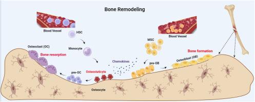
In this study, we constructed a cell atlas encompassing normal, infected, and reconstructed cortical bone. Then, we identified a novel subset at the earlier stage of the osteoclast lineage that exhibited increased expression of IDO1, CCL3, and CCL4. These IDO1highCCL3highCCL4high cells, termed osteostaticytes (OSCs), were further regarded as the reservoir of osteoclasts in the reversal phase. Notably, OSCs exhibited the highest chemotactic activity, surpassing other lineage subsets. We also discovered that cells at the earlier stage of the osteoclast lineage play a significant role in recruiting mesenchymal stem cells (MSCs). Finally, the data revealed that OSCs might be positively related to the occurrence of bone MSCs and the contribution of bone remodelling.
Conclusion
Collectively, our findings revealed a novel stage (OSC) within the osteoclast lineage, potentially representing elusive bone reversal cells due to its increased chemotactic ability towards MSCs and potential contribution to bone remodelling. This study provides valuable insights into the intricate mechanisms of the reversal phase during bone remodelling and unveils potential therapeutic strategies for diseases associated with bone uncoupling.
Translational potential of this article
This study identified a new subset, referred to as IDO1(plus symbol) CCL3(plus symbol) CCL4(plus symbol) osteostaticytes which displayed the highest chemotactic activity among all osteoclast lineages and may serve as reversal cells in bone remodelling. These findings offer new insights and insights for understanding bone reversal-related diseases and may serve as novel therapeutic targets for conditions such as osteomyelitis and delayed bone healing.
期刊介绍:
The Journal of Orthopaedic Translation (JOT) is the official peer-reviewed, open access journal of the Chinese Speaking Orthopaedic Society (CSOS) and the International Chinese Musculoskeletal Research Society (ICMRS). It is published quarterly, in January, April, July and October, by Elsevier.


 分享
分享
 求助内容:
求助内容: 应助结果提醒方式:
 扫码关注我们
