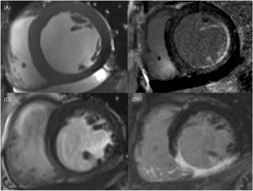
Papillary muscle (PM) infarction (PMI) detected by cardiac magnetic resonance imaging (CMR) is associated with poor outcomes. Whether PM parameters provide more value for mitral regurgitation (MR) management currently remains unclear. Therefore, we examined the prognostic value of PMI using CMR in patients with MR.
Between March 2018 and July 2023, we retrospectively enrolled 397 patients with MR undergoing CMR. CMR was used to detect PMI qualitatively and quantitively. We also collected baseline clinical, echocardiography, and follow-up data.
Of the 397 patients with MR (52.4 ± 13.9 years), 117 (29.5%) were assigned to the PMI group, with 280 (70.5%) in the non-PMI group. PMI was demonstrated more in the posteromedial PM (PM-PM, 98/117) than in the anterolateral PM (AL-PM, 45/117). Compared with patients without PMI, patients with PMI had a decreased AL-PM (41.5 ± 5.4 vs. 45.6 ± 5.3)/PM-PM diastolic length (35.0 ± 5.2 vs. 37.9 ± 4.0), PM-longitudinal strain (LS, 20.4 ± 6.1 vs. 24.9 ± 4.6), AL-PM-LS (19.7 ± 6.8 vs. 24.7 ± 5.6)/PM-PM-LS (21.2 ± 7.9 vs. 25.2 ± 6.0), and increased inter-PM distance (25.7 ± 8.0 vs. 22.7 ± 6.2, all p < 0.001). Multiple logistic regression analyses identified male sex (odds ratio [OR] = 3.65, 95% confidence interval = 1.881−7.081, p < 0.001) diabetes mellitus (OR/95% CI/p = 2.534/1.13–5.68/0.024), AL-PM diastolic length (OR/95% CI/p = 0.841/0.77–0.92/< 0.001), PM-PM diastolic length (OR/95% CI/p = 0.873/0.79–0.964/0.007), inter-PM distance (OR/95% CI/p = 1.087/1.028–1.15/0.003), AL-PM-LS (OR/95% CI/p = 0.892/0.843–0.94/< 0.001), and PM-PM-LS (OR/95% CI/p = 0.95/0.9–0.992/0.021) as independently associated with PMI. Over a 769 ± 367-day follow-up, 100 (25.2%) patients had arrhythmia. Cox regression analyses indicated that PMI (hazard ratio [HR]/95% CI/p = 1.644/1.062–2.547/0.026), AL-PM-LS (HR/95% CI/p = 0.937/0.903–0.973/0.001), and PM-PM-LS (HR/95% CI/p = 0.933/0.902–0.965/< 0.001) remained independently associated with MR.
The CMR-derived PMI and LS parameters improve the evaluation of PM dysfunction, indicating a high risk for arrhythmia, and provide additive risk stratification for patients with MR.


