Sabine Wagner, Christian Ewald, Diana Freitag, Karl-Heinz Herrmann, Arend Koch, Johannes Bauer, Thomas J Vogl, André Kemmling, Hubert Gufler
{"title":"Radiomics and visual analysis for predicting success of transplantation of heterotopic glioblastoma in mice with MRI.","authors":"Sabine Wagner, Christian Ewald, Diana Freitag, Karl-Heinz Herrmann, Arend Koch, Johannes Bauer, Thomas J Vogl, André Kemmling, Hubert Gufler","doi":"10.1007/s11060-024-04725-z","DOIUrl":null,"url":null,"abstract":"<p><strong>Background: </strong>Quantifying tumor growth and treatment response noninvasively poses a challenge to all experimental tumor models. The aim of our study was, to assess the value of quantitative and visual examination and radiomic feature analysis of high-resolution MR images of heterotopic glioblastoma xenografts in mice to determine tumor cell proliferation (TCP).</p><p><strong>Methods: </strong>Human glioblastoma cells were injected subcutaneously into both flanks of immunodeficient mice and followed up on a 3 T MR scanner. Volumes and signal intensities were calculated. Visual assessment of the internal tumor structure was based on a scoring system. Radiomic feature analysis was performed using MaZda software. The results were correlated with histopathology and immunochemistry.</p><p><strong>Results: </strong>21 tumors in 14 animals were analyzed. The volumes of xenografts with high TCP (H-TCP) increased, whereas those with low TCP (L-TCP) or no TCP (N-TCP) continued to decrease over time (p < 0.05). A low intensity rim (rim sign) on unenhanced T1-weighted images provided the highest diagnostic accuracy at visual analysis for assessing H-TCP (p < 0.05). Applying radiomic feature analysis, wavelet transform parameters were best for distinguishing between H-TCP and L-TCP / N-TCP (p < 0.05).</p><p><strong>Conclusion: </strong>Visual and radiomic feature analysis of the internal structure of heterotopically implanted glioblastomas provide reproducible and quantifiable results to predict the success of transplantation.</p>","PeriodicalId":16425,"journal":{"name":"Journal of Neuro-Oncology","volume":" ","pages":"257-267"},"PeriodicalIF":3.1000,"publicationDate":"2024-09-01","publicationTypes":"Journal Article","fieldsOfStudy":null,"isOpenAccess":false,"openAccessPdf":"https://www.ncbi.nlm.nih.gov/pmc/articles/PMC11341603/pdf/","citationCount":"0","resultStr":null,"platform":"Semanticscholar","paperid":null,"PeriodicalName":"Journal of Neuro-Oncology","FirstCategoryId":"3","ListUrlMain":"https://doi.org/10.1007/s11060-024-04725-z","RegionNum":2,"RegionCategory":"医学","ArticlePicture":[],"TitleCN":null,"AbstractTextCN":null,"PMCID":null,"EPubDate":"2024/7/3 0:00:00","PubModel":"Epub","JCR":"Q2","JCRName":"CLINICAL NEUROLOGY","Score":null,"Total":0}
引用次数: 0
Abstract
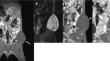
Background: Quantifying tumor growth and treatment response noninvasively poses a challenge to all experimental tumor models. The aim of our study was, to assess the value of quantitative and visual examination and radiomic feature analysis of high-resolution MR images of heterotopic glioblastoma xenografts in mice to determine tumor cell proliferation (TCP).
Methods: Human glioblastoma cells were injected subcutaneously into both flanks of immunodeficient mice and followed up on a 3 T MR scanner. Volumes and signal intensities were calculated. Visual assessment of the internal tumor structure was based on a scoring system. Radiomic feature analysis was performed using MaZda software. The results were correlated with histopathology and immunochemistry.
Results: 21 tumors in 14 animals were analyzed. The volumes of xenografts with high TCP (H-TCP) increased, whereas those with low TCP (L-TCP) or no TCP (N-TCP) continued to decrease over time (p < 0.05). A low intensity rim (rim sign) on unenhanced T1-weighted images provided the highest diagnostic accuracy at visual analysis for assessing H-TCP (p < 0.05). Applying radiomic feature analysis, wavelet transform parameters were best for distinguishing between H-TCP and L-TCP / N-TCP (p < 0.05).
Conclusion: Visual and radiomic feature analysis of the internal structure of heterotopically implanted glioblastomas provide reproducible and quantifiable results to predict the success of transplantation.
期刊介绍:
The Journal of Neuro-Oncology is a multi-disciplinary journal encompassing basic, applied, and clinical investigations in all research areas as they relate to cancer and the central nervous system. It provides a single forum for communication among neurologists, neurosurgeons, radiotherapists, medical oncologists, neuropathologists, neurodiagnosticians, and laboratory-based oncologists conducting relevant research. The Journal of Neuro-Oncology does not seek to isolate the field, but rather to focus the efforts of many disciplines in one publication through a format which pulls together these diverse interests. More than any other field of oncology, cancer of the central nervous system requires multi-disciplinary approaches. To alleviate having to scan dozens of journals of cell biology, pathology, laboratory and clinical endeavours, JNO is a periodical in which current, high-quality, relevant research in all aspects of neuro-oncology may be found.


 分享
分享
 求助内容:
求助内容: 应助结果提醒方式:
 扫码关注我们
