{"title":"Brain structure alterations following neonatal exposure to low-frequency electromagnetic fields: A histological analysis","authors":"Stephanie M. Sissons, Blake T. Dotta","doi":"10.1002/jdn.10361","DOIUrl":null,"url":null,"abstract":"<p>Nitric oxide (NO) and electromagnetic fields (EMF) have been extensively studied for their roles in neurobiology, particularly in regulating cerebral functions and synaptic plasticity. This study investigates the impact of EMFs on NO modulation and its subsequent effects on neurodevelopment, building upon prior research examining EMF exposure's consequences on Wistar albino rats. Rats were exposed perinatally to either tap water, 1 g/L of L-arginine (LA) or 0.5 g/L of <i>N</i>-methylarginine (NMA). Half of the rats in each group were also exposed to a 7-Hz square-wave EMF at three separate intensities (5, 50 and 500 nT) for 2–14 days following birth. Animals were allowed to develop, and their brains were harvested later in adulthood (mean age = 568.17 days, SD = 162.73). Histological analyses were used to elucidate structural changes in key brain regions. All brains were stained with Toluidine Blue O (TBO), enabling the visualization of neurons. Neuronal counts were then conducted in specific regions of interest (e.g. hippocampus, cortices, amygdala and hypothalamus). Histological analyses revealed significant alterations in neuronal density in specific brain regions, particularly in response to EMF exposure and pharmacological interventions. Notable findings include a main EMF exposure effect where increased neuronal counts were observed in the secondary somatosensory cortex under low EMF intensities (<i>p</i> < 0.001) and sex-specific responses in the hippocampus, where a significant increase in neuronal counts was observed in the left CA3 region in female rats exposed to EMF compared to unexposed females (<i>t</i>(18) = 2.371, <i>p</i> = 0.029). Additionally, a significant increase in neuronal counts in the right entorhinal cortex was seen in male rats exposed to EMF compared to unexposed males (<i>t</i>(18) = 2.216, <i>p</i> = 0.040). These findings emphasize the complex interaction among sex, EMF exposure and pharmacological agents on neuronal dynamics across brain regions, highlighting the need for further research to identify underlying mechanisms and potential implications for cognitive function and neurological health in clinical and environmental contexts.</p>","PeriodicalId":13914,"journal":{"name":"International Journal of Developmental Neuroscience","volume":"84 7","pages":"651-658"},"PeriodicalIF":1.6000,"publicationDate":"2024-07-05","publicationTypes":"Journal Article","fieldsOfStudy":null,"isOpenAccess":false,"openAccessPdf":"https://onlinelibrary.wiley.com/doi/epdf/10.1002/jdn.10361","citationCount":"0","resultStr":null,"platform":"Semanticscholar","paperid":null,"PeriodicalName":"International Journal of Developmental Neuroscience","FirstCategoryId":"3","ListUrlMain":"https://onlinelibrary.wiley.com/doi/10.1002/jdn.10361","RegionNum":4,"RegionCategory":"医学","ArticlePicture":[],"TitleCN":null,"AbstractTextCN":null,"PMCID":null,"EPubDate":"","PubModel":"","JCR":"Q3","JCRName":"DEVELOPMENTAL BIOLOGY","Score":null,"Total":0}
引用次数: 0
Abstract
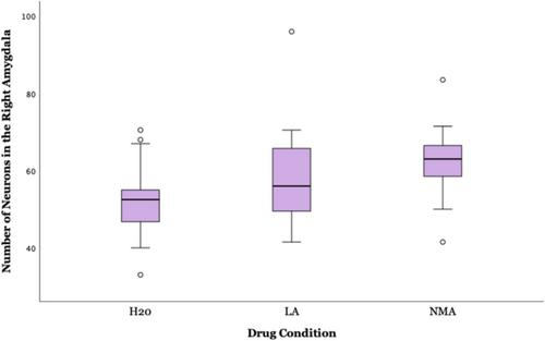
Nitric oxide (NO) and electromagnetic fields (EMF) have been extensively studied for their roles in neurobiology, particularly in regulating cerebral functions and synaptic plasticity. This study investigates the impact of EMFs on NO modulation and its subsequent effects on neurodevelopment, building upon prior research examining EMF exposure's consequences on Wistar albino rats. Rats were exposed perinatally to either tap water, 1 g/L of L-arginine (LA) or 0.5 g/L of N-methylarginine (NMA). Half of the rats in each group were also exposed to a 7-Hz square-wave EMF at three separate intensities (5, 50 and 500 nT) for 2–14 days following birth. Animals were allowed to develop, and their brains were harvested later in adulthood (mean age = 568.17 days, SD = 162.73). Histological analyses were used to elucidate structural changes in key brain regions. All brains were stained with Toluidine Blue O (TBO), enabling the visualization of neurons. Neuronal counts were then conducted in specific regions of interest (e.g. hippocampus, cortices, amygdala and hypothalamus). Histological analyses revealed significant alterations in neuronal density in specific brain regions, particularly in response to EMF exposure and pharmacological interventions. Notable findings include a main EMF exposure effect where increased neuronal counts were observed in the secondary somatosensory cortex under low EMF intensities (p < 0.001) and sex-specific responses in the hippocampus, where a significant increase in neuronal counts was observed in the left CA3 region in female rats exposed to EMF compared to unexposed females (t(18) = 2.371, p = 0.029). Additionally, a significant increase in neuronal counts in the right entorhinal cortex was seen in male rats exposed to EMF compared to unexposed males (t(18) = 2.216, p = 0.040). These findings emphasize the complex interaction among sex, EMF exposure and pharmacological agents on neuronal dynamics across brain regions, highlighting the need for further research to identify underlying mechanisms and potential implications for cognitive function and neurological health in clinical and environmental contexts.
期刊介绍:
International Journal of Developmental Neuroscience publishes original research articles and critical review papers on all fundamental and clinical aspects of nervous system development, renewal and regeneration, as well as on the effects of genetic and environmental perturbations of brain development and homeostasis leading to neurodevelopmental disorders and neurological conditions. Studies describing the involvement of stem cells in nervous system maintenance and disease (including brain tumours), stem cell-based approaches for the investigation of neurodegenerative diseases, roles of neuroinflammation in development and disease, and neuroevolution are also encouraged. Investigations using molecular, cellular, physiological, genetic and epigenetic approaches in model systems ranging from simple invertebrates to human iPSC-based 2D and 3D models are encouraged, as are studies using experimental models that provide behavioural or evolutionary insights. The journal also publishes Special Issues dealing with topics at the cutting edge of research edited by Guest Editors appointed by the Editor in Chief. A major aim of the journal is to facilitate the transfer of fundamental studies of nervous system development, maintenance, and disease to clinical applications. The journal thus intends to disseminate valuable information for both biologists and physicians. International Journal of Developmental Neuroscience is owned and supported by The International Society for Developmental Neuroscience (ISDN), an organization of scientists interested in advancing developmental neuroscience research in the broadest sense.


 分享
分享
 求助内容:
求助内容: 应助结果提醒方式:
 扫码关注我们
