{"title":"Pyogenic granuloma of the hard palate leading to alveolar cleft: a case report.","authors":"Woo Jin Song, Hyun Beom Choi, Min Sung Tak","doi":"10.7181/acfs.2024.00080","DOIUrl":null,"url":null,"abstract":"<p><p>This case report describes a rare occurrence of pyogenic granuloma (PG) in the hard palate deviating from its typical gingival location that led to the formation of an alveolar cleft. The aggressive growth pattern of the lesion, with atypical progression from a pedunculated nodule to an alveolar cleft, raised concern. The diagnosis was based on magnetic resonance imaging and computed tomography findings, which revealed a tadpole-shaped lesion originating from the midline hard palate. The differential diagnosis included a minor salivary gland tumor. Surgical excision was performed under general anesthesia and resulted in a mucosal defect without nasolabial fistula formation or bone exposure. The palatal defect was packed with oxidized regenerated cellulose and closed with Vicryl Rapide sutures, both of which contributed to the patient's successful outcomes. Our comprehensive approach, extending across the stages of surgical planning, execution, and postoperative care, demonstrated the advantages of a multidisciplinary strategy for the accurate diagnosis and effective treatment of palatal PGs. This report makes a meaningful contribution to the existing literature on common oral lesions by emphasizing the importance of a broad differential diagnosis and a systematic approach to oral pathologies. It also raises clinical awareness of PGs with atypical presentations and the diagnostic challenge that they pose.</p>","PeriodicalId":52238,"journal":{"name":"Archives of Craniofacial Surgery","volume":"25 3","pages":"150-154"},"PeriodicalIF":0.0000,"publicationDate":"2024-06-01","publicationTypes":"Journal Article","fieldsOfStudy":null,"isOpenAccess":false,"openAccessPdf":"https://www.ncbi.nlm.nih.gov/pmc/articles/PMC11231411/pdf/","citationCount":"0","resultStr":null,"platform":"Semanticscholar","paperid":null,"PeriodicalName":"Archives of Craniofacial Surgery","FirstCategoryId":"1085","ListUrlMain":"https://doi.org/10.7181/acfs.2024.00080","RegionNum":0,"RegionCategory":null,"ArticlePicture":[],"TitleCN":null,"AbstractTextCN":null,"PMCID":null,"EPubDate":"2024/6/20 0:00:00","PubModel":"Epub","JCR":"Q2","JCRName":"Medicine","Score":null,"Total":0}
引用次数: 0
Abstract
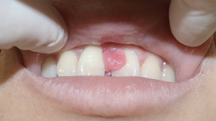
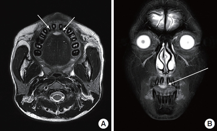
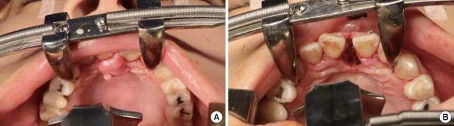
This case report describes a rare occurrence of pyogenic granuloma (PG) in the hard palate deviating from its typical gingival location that led to the formation of an alveolar cleft. The aggressive growth pattern of the lesion, with atypical progression from a pedunculated nodule to an alveolar cleft, raised concern. The diagnosis was based on magnetic resonance imaging and computed tomography findings, which revealed a tadpole-shaped lesion originating from the midline hard palate. The differential diagnosis included a minor salivary gland tumor. Surgical excision was performed under general anesthesia and resulted in a mucosal defect without nasolabial fistula formation or bone exposure. The palatal defect was packed with oxidized regenerated cellulose and closed with Vicryl Rapide sutures, both of which contributed to the patient's successful outcomes. Our comprehensive approach, extending across the stages of surgical planning, execution, and postoperative care, demonstrated the advantages of a multidisciplinary strategy for the accurate diagnosis and effective treatment of palatal PGs. This report makes a meaningful contribution to the existing literature on common oral lesions by emphasizing the importance of a broad differential diagnosis and a systematic approach to oral pathologies. It also raises clinical awareness of PGs with atypical presentations and the diagnostic challenge that they pose.
本病例报告描述了一起罕见的硬腭化脓性肉芽肿(PG)病例,该病例偏离了其典型的牙龈位置,导致牙槽裂的形成。病变的侵袭性生长模式,以及从蒂状结节到齿槽裂的非典型进展,引起了人们的关注。诊断依据是磁共振成像和计算机断层扫描结果,结果显示病变呈蝌蚪状,起源于硬腭中线。鉴别诊断包括轻微的唾液腺肿瘤。手术切除是在全身麻醉的情况下进行的,术后出现了粘膜缺损,但没有形成鼻唇瘘或骨头暴露。腭部缺损处用氧化再生纤维素填塞,并用 Vicryl Rapide 缝线缝合,这两种方法都有助于患者取得成功。我们的综合方法贯穿了手术计划、实施和术后护理的各个阶段,证明了多学科策略在准确诊断和有效治疗腭PG方面的优势。本报告强调了广泛鉴别诊断和系统治疗口腔病变的重要性,为现有的常见口腔病变文献做出了有意义的贡献。它还提高了临床对表现不典型的腭咽癌及其诊断挑战的认识。


 分享
分享
 求助内容:
求助内容: 应助结果提醒方式:
 扫码关注我们
