Evaluation of the proximal femur using the digital photographs: Does change in proximal femur position due to anteversion affect the measurement of the size of the femoral head diameter?
Olasode Israel Akinmokun, Utibeabasi Ime Edem, Olanrewaju Matthew Adeoye
{"title":"Evaluation of the proximal femur using the digital photographs: Does change in proximal femur position due to anteversion affect the measurement of the size of the femoral head diameter?","authors":"Olasode Israel Akinmokun, Utibeabasi Ime Edem, Olanrewaju Matthew Adeoye","doi":"10.4103/jwas.jwas_145_23","DOIUrl":null,"url":null,"abstract":"<p><strong>Background: </strong>A plain pelvic radiograph is usually conducted with the lower limbs in internal rotation. This is to correct the anteversion of the femur. However, in the fracture neck of the femur, internal rotation of the fractured limb is avoided, because it would be painful. We examined the effect of correction of anteversion or otherwise on the diameter of the head of the femur using imaging.</p><p><strong>Objectives: </strong>This study aimed to determine if there was a significant difference between the femoral head diameter at two different positions, at the normal anatomical position (without correcting the anteversion) and at the corrected anteversion position. It also aimed to document the correlation and the statistical significance between the differences in the size of the diameter at these two different positions with the anteversion angles of the femoral bone.</p><p><strong>Materials and methods: </strong>Two sets of digital photographs of the proximal part of 55 non-sexed, non-paired femoral bones were taken. Images obtained were at two positions: normal anatomical (with anteversion uncorrected) and anteversion corrected positions. The diameters of the head of the femur were documented at these two different positions. The anteversion angles and actual femoral head (AFH) diameters were also measured and documented.</p><p><strong>Results: </strong>The femoral head diameters at anatomical positions were persistently larger than those measured after the anteversion was corrected, except in three femoral bones (5%) where no differences were observed. The difference in the two measurements was statistically significant to the anteversion angle of the femoral bone. (<i>P</i> = 0.0005). The means of the two sets of measurements were statistically different from each other. Pairwise correlation showed that both were strongly associated with the AFH diameter but the measurements from images with corrected anteversion had a higher value (0.8166) than the measurements from normal anatomical position (0.7526).</p><p><strong>Conclusion: </strong>The correction of femoral anteversion produced femoral head size measurements that were closer to AFH diameters compared to those without the correction of the femoral anteversion. Femoral anteversion should always be corrected as per protocol.</p>","PeriodicalId":73993,"journal":{"name":"Journal of the West African College of Surgeons","volume":"14 3","pages":"314-318"},"PeriodicalIF":0.0000,"publicationDate":"2024-07-01","publicationTypes":"Journal Article","fieldsOfStudy":null,"isOpenAccess":false,"openAccessPdf":"https://www.ncbi.nlm.nih.gov/pmc/articles/PMC11232775/pdf/","citationCount":"0","resultStr":null,"platform":"Semanticscholar","paperid":null,"PeriodicalName":"Journal of the West African College of Surgeons","FirstCategoryId":"1085","ListUrlMain":"https://doi.org/10.4103/jwas.jwas_145_23","RegionNum":0,"RegionCategory":null,"ArticlePicture":[],"TitleCN":null,"AbstractTextCN":null,"PMCID":null,"EPubDate":"2024/5/24 0:00:00","PubModel":"Epub","JCR":"","JCRName":"","Score":null,"Total":0}
引用次数: 0
Abstract
Background: A plain pelvic radiograph is usually conducted with the lower limbs in internal rotation. This is to correct the anteversion of the femur. However, in the fracture neck of the femur, internal rotation of the fractured limb is avoided, because it would be painful. We examined the effect of correction of anteversion or otherwise on the diameter of the head of the femur using imaging.
Objectives: This study aimed to determine if there was a significant difference between the femoral head diameter at two different positions, at the normal anatomical position (without correcting the anteversion) and at the corrected anteversion position. It also aimed to document the correlation and the statistical significance between the differences in the size of the diameter at these two different positions with the anteversion angles of the femoral bone.
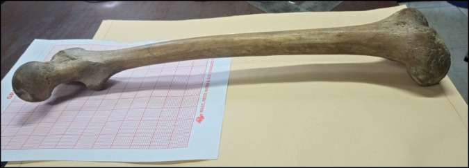
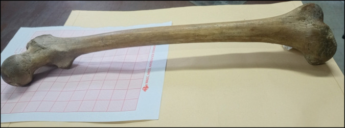
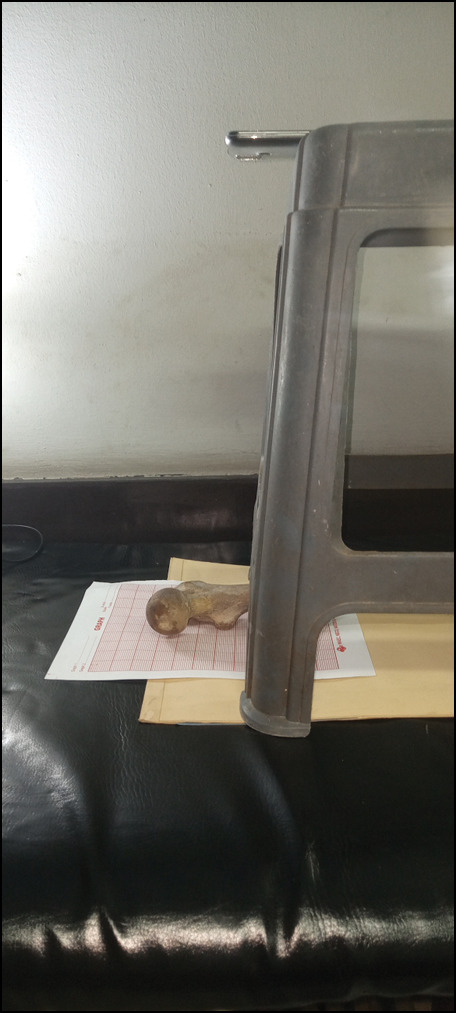
Materials and methods: Two sets of digital photographs of the proximal part of 55 non-sexed, non-paired femoral bones were taken. Images obtained were at two positions: normal anatomical (with anteversion uncorrected) and anteversion corrected positions. The diameters of the head of the femur were documented at these two different positions. The anteversion angles and actual femoral head (AFH) diameters were also measured and documented.
Results: The femoral head diameters at anatomical positions were persistently larger than those measured after the anteversion was corrected, except in three femoral bones (5%) where no differences were observed. The difference in the two measurements was statistically significant to the anteversion angle of the femoral bone. (P = 0.0005). The means of the two sets of measurements were statistically different from each other. Pairwise correlation showed that both were strongly associated with the AFH diameter but the measurements from images with corrected anteversion had a higher value (0.8166) than the measurements from normal anatomical position (0.7526).
Conclusion: The correction of femoral anteversion produced femoral head size measurements that were closer to AFH diameters compared to those without the correction of the femoral anteversion. Femoral anteversion should always be corrected as per protocol.


 分享
分享

 求助内容:
求助内容: 应助结果提醒方式:
 扫码关注我们
