Radiomics and machine learning analysis of liver magnetic resonance imaging for prediction and early detection of tumor response in colorectal liver metastases.
Sungjin Yoon, Young Jae Kim, Ji Soo Jeon, Su Joa Ahn, Seung Joon Choi
{"title":"Radiomics and machine learning analysis of liver magnetic resonance imaging for prediction and early detection of tumor response in colorectal liver metastases.","authors":"Sungjin Yoon, Young Jae Kim, Ji Soo Jeon, Su Joa Ahn, Seung Joon Choi","doi":"10.14216/kjco.24005","DOIUrl":null,"url":null,"abstract":"<p><strong>Purpose: </strong>The aim of this study was to demonstrate the effectiveness of a machine learning-based radiomics model for distinguishing tumor response and overall survival in patients with unresectable colorectal liver metastases (CRLM) treated with targeted biological therapy.</p><p><strong>Methods: </strong>We prospectively recruited 17 patients with unresectable liver metastases of colorectal cancer, who had been given targeted biological therapy as the first line of treatment. All patients underwent liver magnetic resonance imaging (MRI) three times up until 8 weeks after chemotherapy. We evaluated the diagnostic performance of machine learning-based radiomics model in tumor response of liver MRI compared with the guidelines for the Response Evaluation Criteria in Solid Tumors. Overall survival was evaluated using the Kaplan-Meier analysis and compared to the Cox proportional hazard ratios following univariate and multivariate analyses.</p><p><strong>Results: </strong>Performance measurement of the trained model through metrics showed the accuracy of the machine learning model to be 76.5%, and the area under the receiver operating characteristic curve was 0.857 (95% confidence interval [CI], 0.605-0.976; P < 0.001). For the patients classified as non-progressing or progressing by the radiomics model, the median overall survival was 17.5 months (95% CI, 12.8-22.2), and 14.8 months (95% CI, 14.2-15.4), respectively (P = 0.431, log-rank test).</p><p><strong>Conclusion: </strong>Machine learning-based radiomics models could have the potential to predict tumor response in patients with unresectable CRLM treated with biologic therapy.</p>","PeriodicalId":74045,"journal":{"name":"Korean journal of clinical oncology","volume":"20 1","pages":"27-35"},"PeriodicalIF":0.0000,"publicationDate":"2024-05-01","publicationTypes":"Journal Article","fieldsOfStudy":null,"isOpenAccess":false,"openAccessPdf":"https://www.ncbi.nlm.nih.gov/pmc/articles/PMC11261177/pdf/","citationCount":"0","resultStr":null,"platform":"Semanticscholar","paperid":null,"PeriodicalName":"Korean journal of clinical oncology","FirstCategoryId":"1085","ListUrlMain":"https://doi.org/10.14216/kjco.24005","RegionNum":0,"RegionCategory":null,"ArticlePicture":[],"TitleCN":null,"AbstractTextCN":null,"PMCID":null,"EPubDate":"2024/6/30 0:00:00","PubModel":"Epub","JCR":"","JCRName":"","Score":null,"Total":0}
引用次数: 0
Abstract
Purpose: The aim of this study was to demonstrate the effectiveness of a machine learning-based radiomics model for distinguishing tumor response and overall survival in patients with unresectable colorectal liver metastases (CRLM) treated with targeted biological therapy.
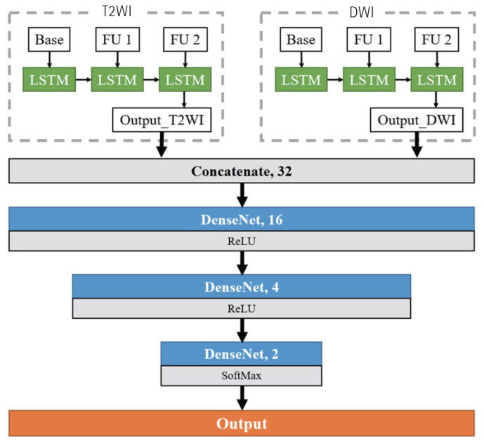
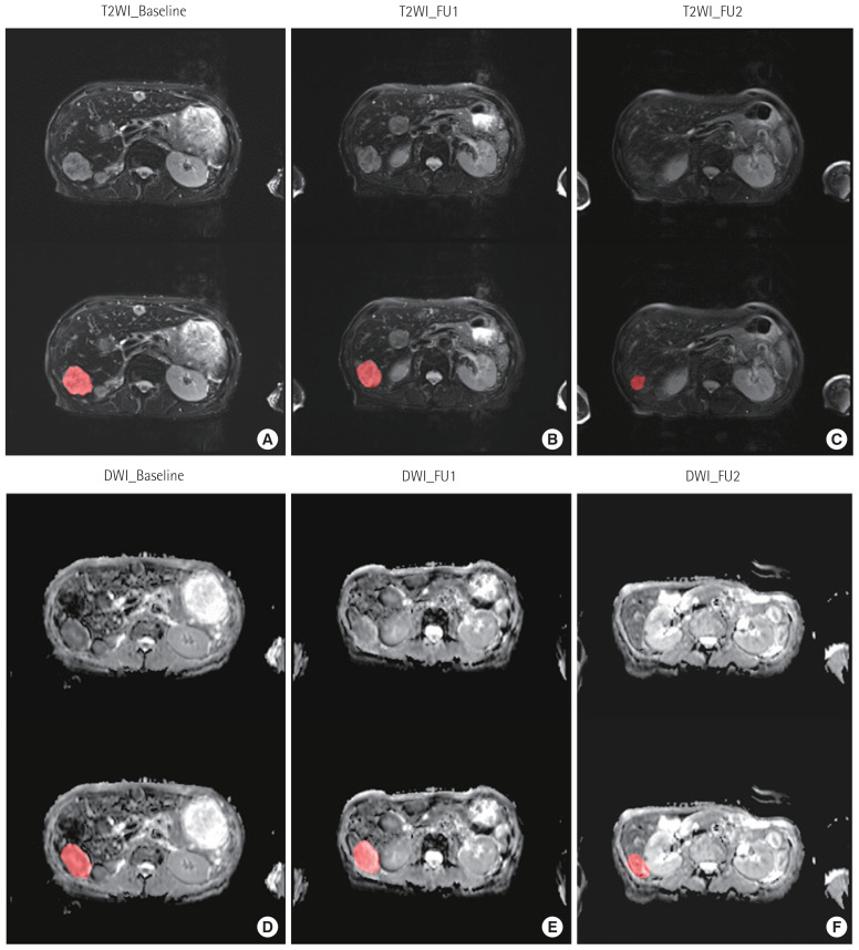
Methods: We prospectively recruited 17 patients with unresectable liver metastases of colorectal cancer, who had been given targeted biological therapy as the first line of treatment. All patients underwent liver magnetic resonance imaging (MRI) three times up until 8 weeks after chemotherapy. We evaluated the diagnostic performance of machine learning-based radiomics model in tumor response of liver MRI compared with the guidelines for the Response Evaluation Criteria in Solid Tumors. Overall survival was evaluated using the Kaplan-Meier analysis and compared to the Cox proportional hazard ratios following univariate and multivariate analyses.
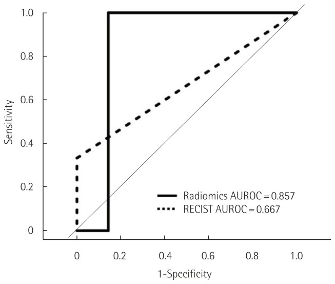
Results: Performance measurement of the trained model through metrics showed the accuracy of the machine learning model to be 76.5%, and the area under the receiver operating characteristic curve was 0.857 (95% confidence interval [CI], 0.605-0.976; P < 0.001). For the patients classified as non-progressing or progressing by the radiomics model, the median overall survival was 17.5 months (95% CI, 12.8-22.2), and 14.8 months (95% CI, 14.2-15.4), respectively (P = 0.431, log-rank test).
Conclusion: Machine learning-based radiomics models could have the potential to predict tumor response in patients with unresectable CRLM treated with biologic therapy.


 分享
分享

 求助内容:
求助内容: 应助结果提醒方式:
 扫码关注我们
