Samuel L. Rose , Felix Martín Ferroni , Sam Horrell , Carlos Dante Brondino , Robert R. Eady , Sofia Jaho , Michael A. Hough , Robin L. Owen , Svetlana V. Antonyuk , S. Samar Hasnain
{"title":"Spectroscopically Validated pH-dependent MSOX Movies Provide Detailed Mechanism of Copper Nitrite Reductases","authors":"Samuel L. Rose , Felix Martín Ferroni , Sam Horrell , Carlos Dante Brondino , Robert R. Eady , Sofia Jaho , Michael A. Hough , Robin L. Owen , Svetlana V. Antonyuk , S. Samar Hasnain","doi":"10.1016/j.jmb.2024.168706","DOIUrl":null,"url":null,"abstract":"<div><p>Copper nitrite reductases (CuNiRs) exhibit a strong pH dependence of their catalytic activity. Structural movies can be obtained by serially recording multiple structures (frames) from the same spot of a crystal using the MSOX serial crystallography approach. This method has been combined with on-line single crystal optical spectroscopy to capture the pH-dependent structural changes that accompany during turnover of CuNiRs from two <em>Rhizobia</em> species. The structural movies, initiated by the redox activation of a type-1 copper site (T1Cu) <em>via</em> X-ray generated photoelectrons, have been obtained for the substrate-free and substrate-bound states at low (high enzymatic activity) and high (low enzymatic activity) pH. At low pH, formation of the product nitric oxide (NO) is complete at the catalytic type-2 copper site (T2Cu) after a dose of 3 MGy (frame 5) with full bleaching of the T1Cu ligand-to-metal charge transfer (LMCT) 455 nm band (S(σ)<sub>Cys</sub> → T1Cu<sup>2+</sup>) which in itself indicates the electronic route of proton-coupled electron transfer (PCET) from T1Cu to T2Cu. In contrast at high pH, the changes in optical spectra are relatively small and the formation of NO is only observed in later frames (frame 15 in <em>Br<sup>2D</sup></em>NiR, 10 MGy), consistent with the loss of PCET required for catalysis. This is accompanied by decarboxylation of the catalytic Asp<sub>CAT</sub> residue, with CO<sub>2</sub> trapped in the catalytic pocket.</p></div>","PeriodicalId":369,"journal":{"name":"Journal of Molecular Biology","volume":"436 18","pages":"Article 168706"},"PeriodicalIF":4.5000,"publicationDate":"2024-09-15","publicationTypes":"Journal Article","fieldsOfStudy":null,"isOpenAccess":false,"openAccessPdf":"https://www.sciencedirect.com/science/article/pii/S0022283624003152/pdfft?md5=6b7fe5e7cd5a1c41f3fbd91a48baf02b&pid=1-s2.0-S0022283624003152-main.pdf","citationCount":"0","resultStr":null,"platform":"Semanticscholar","paperid":null,"PeriodicalName":"Journal of Molecular Biology","FirstCategoryId":"99","ListUrlMain":"https://www.sciencedirect.com/science/article/pii/S0022283624003152","RegionNum":2,"RegionCategory":"生物学","ArticlePicture":[],"TitleCN":null,"AbstractTextCN":null,"PMCID":null,"EPubDate":"2024/7/14 0:00:00","PubModel":"Epub","JCR":"Q1","JCRName":"BIOCHEMISTRY & MOLECULAR BIOLOGY","Score":null,"Total":0}
引用次数: 0
Abstract
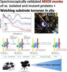
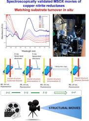
Copper nitrite reductases (CuNiRs) exhibit a strong pH dependence of their catalytic activity. Structural movies can be obtained by serially recording multiple structures (frames) from the same spot of a crystal using the MSOX serial crystallography approach. This method has been combined with on-line single crystal optical spectroscopy to capture the pH-dependent structural changes that accompany during turnover of CuNiRs from two Rhizobia species. The structural movies, initiated by the redox activation of a type-1 copper site (T1Cu) via X-ray generated photoelectrons, have been obtained for the substrate-free and substrate-bound states at low (high enzymatic activity) and high (low enzymatic activity) pH. At low pH, formation of the product nitric oxide (NO) is complete at the catalytic type-2 copper site (T2Cu) after a dose of 3 MGy (frame 5) with full bleaching of the T1Cu ligand-to-metal charge transfer (LMCT) 455 nm band (S(σ)Cys → T1Cu2+) which in itself indicates the electronic route of proton-coupled electron transfer (PCET) from T1Cu to T2Cu. In contrast at high pH, the changes in optical spectra are relatively small and the formation of NO is only observed in later frames (frame 15 in Br2DNiR, 10 MGy), consistent with the loss of PCET required for catalysis. This is accompanied by decarboxylation of the catalytic AspCAT residue, with CO2 trapped in the catalytic pocket.
期刊介绍:
Journal of Molecular Biology (JMB) provides high quality, comprehensive and broad coverage in all areas of molecular biology. The journal publishes original scientific research papers that provide mechanistic and functional insights and report a significant advance to the field. The journal encourages the submission of multidisciplinary studies that use complementary experimental and computational approaches to address challenging biological questions.
Research areas include but are not limited to: Biomolecular interactions, signaling networks, systems biology; Cell cycle, cell growth, cell differentiation; Cell death, autophagy; Cell signaling and regulation; Chemical biology; Computational biology, in combination with experimental studies; DNA replication, repair, and recombination; Development, regenerative biology, mechanistic and functional studies of stem cells; Epigenetics, chromatin structure and function; Gene expression; Membrane processes, cell surface proteins and cell-cell interactions; Methodological advances, both experimental and theoretical, including databases; Microbiology, virology, and interactions with the host or environment; Microbiota mechanistic and functional studies; Nuclear organization; Post-translational modifications, proteomics; Processing and function of biologically important macromolecules and complexes; Molecular basis of disease; RNA processing, structure and functions of non-coding RNAs, transcription; Sorting, spatiotemporal organization, trafficking; Structural biology; Synthetic biology; Translation, protein folding, chaperones, protein degradation and quality control.


 分享
分享
 求助内容:
求助内容: 应助结果提醒方式:
 扫码关注我们
