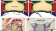
{"title":"How I do it: Endoscopic endonasal resection of the medial wall of the cavernous sinus.","authors":"Eugenio Cárdenas Ruiz-Valdepeñas, Estrella Barrero Ruiz, Aberto Acitores Cancela, Victor Rodriguez Berrocal","doi":"10.1007/s00701-024-06177-w","DOIUrl":null,"url":null,"abstract":"<p><strong>Background: </strong>Resection of the medial wall of the cavernous sinus (MWCSR) is a growing surgical maneuver for the radical removal of pituitary adenomas.</p><p><strong>Method: </strong>We present a simple modification of the technique following the two dural layers of the floor of the sella turcica, allowing for early identification of the medial wall and simplifying dissection. We support this technique with an anatomical analysis on cadaveric specimens and clarifying dissection images.</p><p><strong>Conclusion: </strong>Recognition and dissection of the dural unfolding of the floor of the sella turcica are \"key points\" that lower the risk and facilitate the MWCSR.</p>","PeriodicalId":7370,"journal":{"name":"Acta Neurochirurgica","volume":"166 1","pages":"298"},"PeriodicalIF":1.9000,"publicationDate":"2024-07-16","publicationTypes":"Journal Article","fieldsOfStudy":null,"isOpenAccess":false,"openAccessPdf":"","citationCount":"0","resultStr":null,"platform":"Semanticscholar","paperid":null,"PeriodicalName":"Acta Neurochirurgica","FirstCategoryId":"3","ListUrlMain":"https://doi.org/10.1007/s00701-024-06177-w","RegionNum":3,"RegionCategory":"医学","ArticlePicture":[],"TitleCN":null,"AbstractTextCN":null,"PMCID":null,"EPubDate":"","PubModel":"","JCR":"Q3","JCRName":"CLINICAL NEUROLOGY","Score":null,"Total":0}
引用次数: 0
Abstract
Background: Resection of the medial wall of the cavernous sinus (MWCSR) is a growing surgical maneuver for the radical removal of pituitary adenomas.
Method: We present a simple modification of the technique following the two dural layers of the floor of the sella turcica, allowing for early identification of the medial wall and simplifying dissection. We support this technique with an anatomical analysis on cadaveric specimens and clarifying dissection images.
Conclusion: Recognition and dissection of the dural unfolding of the floor of the sella turcica are "key points" that lower the risk and facilitate the MWCSR.
期刊介绍:
The journal "Acta Neurochirurgica" publishes only original papers useful both to research and clinical work. Papers should deal with clinical neurosurgery - diagnosis and diagnostic techniques, operative surgery and results, postoperative treatment - or with research work in neuroscience if the underlying questions or the results are of neurosurgical interest. Reports on congresses are given in brief accounts. As official organ of the European Association of Neurosurgical Societies the journal publishes all announcements of the E.A.N.S. and reports on the activities of its member societies. Only contributions written in English will be accepted.


 分享
分享
 求助内容:
求助内容: 应助结果提醒方式:
 扫码关注我们
